

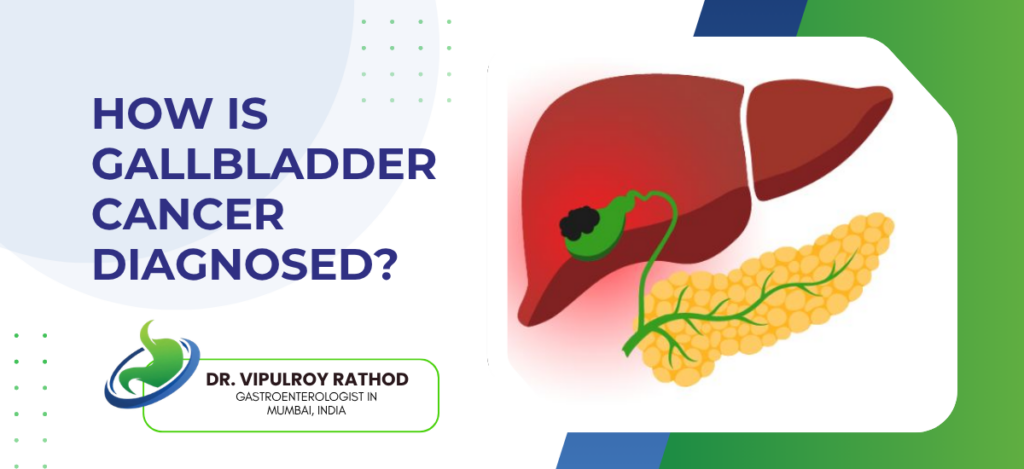
Gallbladder cancer is diagnosed through a sequence of imaging that starts with ultrasound and escalates to CT, MRI, and EUS depending on what’s found. The difficulty is that early disease produces no specific symptoms, so a large share of cases turn up incidentally, either on a scan done for something else or on histology after a routine gallbladder removal. Accurate staging once a lesion is identified is what determines whether treatment is possible, which makes the diagnostic pathway as important as the finding itself.
According to Dr. Vipulroy Rathod, Gastroenterologist in Mumbai, “Gallbladder cancer is one of the hardest GI cancers to catch early because the gallbladder doesn’t announce trouble until late. When a suspicious thickening or mass shows up, the priority is accurate staging, and that’s where EUS adds detail that a standard scan simply can’t provide.”
What Tests Are Used to Diagnose Gallbladder Cancer?
Diagnosis builds in steps, each test answering a question the previous one couldn’t.
Ultrasound comes first, picking up gallbladder wall thickening, masses, or polyps that warrant further investigation, though it can’t confirm cancer on its own.
CT and MRI map the extent, showing whether a tumour has spread to the liver, bile ducts, or distant sites, which is essential for staging once ultrasound raises suspicion.
EUS adds the detail external scans miss, assessing tumour depth and nearby lymph nodes from inside the GI tract at a proximity no surface imaging can reach.
When tissue confirmation is needed, EUS-guided fine needle aspiration samples the lesion directly, settling an ambiguous imaging picture without surgery.
Getting this sequence right requires a gallbladder cancer specialist who can interpret each step and decide what the next investigation should be.
Why Is Gallbladder Cancer So Often Found Late?
The diagnostic challenge isn’t usually the tests themselves but how late the suspicion arises.
No specific early symptoms exist. The vague right upper abdominal discomfort that gallbladder cancer produces is identical to ordinary gallstone disease, so it rarely triggers cancer-focused investigation.
Many cancers surface incidentally on histology after a gallbladder is removed for gallstones, meaning the diagnosis arrives only once the organ is already out.
Imaging overlap muddies the picture, because gallstones and chronic inflammation can mask or mimic an early tumour on ultrasound, delaying the escalation to CT or EUS.
High-risk patients often go unmonitored, as those with large gallstones or gallbladder polyps frequently aren’t placed on the surveillance that would catch a developing cancer early.
For patients dealing with gallbladder-related complications, understanding how endoscopic diagnosis works in the biliary system is a useful background. Read more on bile leakage to see how endoscopic techniques diagnose and manage biliary complications after gallbladder surgery.
Why Choose Dr. Vipulroy Rathod for Gallbladder Cancer?
Dr. Vipulroy Rathod has been diagnosing hepatobiliary cancers at Fortis Hospital Mulund for over 30 years, and his EUS practice since 1998 gives him the staging tool most centres don’t deploy for gallbladder lesions that surface imaging can’t fully characterise. Patients with an ambiguous gallbladder finding often get a clear diagnosis and accurate stage in a single workup rather than a series of scattered scans. Physicians from 35 countries have trained under his approach at Fortis.
Book your consultation today with one of India’s most experienced specialists for gallbladder cancer diagnosis and endoscopic staging.
Frequently Asked Questions
Gallbladder cancer is diagnosed through ultrasound as the first step, followed by CT or MRI for staging, and EUS with fine needle aspiration for tissue confirmation and assessment of local spread.
EUS provides the most accurate local staging by assessing tumour depth and lymph node involvement, and allows fine needle aspiration for tissue diagnosis when imaging findings are inconclusive.
Ultrasound can detect gallbladder wall thickening, masses, or polyps suggestive of cancer, but further imaging with CT, MRI, or EUS is needed to confirm the diagnosis and stage the disease.
Yes. Many gallbladder cancers are found incidentally on histology after gallbladder removal for gallstones, or on imaging performed for an unrelated reason, rather than through symptoms.
Disclaimer:
This blog is intended for informational and educational purposes only and should not be considered medical advice.
Please consult a qualified gastroenterologist or healthcare professional for personalized diagnosis and treatment.
