Dr. Vipulroy Rathod

Author name: Dr. Rathod Medical Foundation

Polyp Found During Colonoscopy: What Next

Most polyps found during colonoscopy are removed right there during the same procedure. The doctor uses a wire loop or forceps through the scope to snip the polyp off the colon wall and sends it to a lab for biopsy. What the biopsy report says about that polyp, its type, size, and whether it shows any precancerous changes, is what decides everything that follows including how soon you need your next colonoscopy. According to Dr. Vipulroy Rathod, an experienced Gastroenterologist in Mumbai, “Finding a polyp during colonoscopy isn’t bad news by itself, it’s actually the system working exactly the way it should because catching polyps early and removing them on the spot is the whole reason screening exists in the first place.” What happens to the polyp after removal? The removal part is actually the easy bit. What matters more is what the lab says about that polyp once it’s been sliced, stained and put under a microscope, because that report is what decides everything going forward. Pathology report: Every polyp goes to a pathologist. They cut it open, look at cell patterns, check for dysplasia. The report takes about 5 to 7 working days to come back. Your gastroenterologist reads it before sitting down with you to discuss what it means. Nobody should be telling you next steps before that report is in hand. Polyp type: This is the part that actually determines risk. Adenomatous polyps are the ones that can eventually go cancerous if left alone long enough. Hyperplastic polyps are almost always harmless. Sessile serrated polyps are a bit of a grey area. The type written on that pathology report sets the tone for your entire follow-up plan. Size: A tiny 3 mm polyp and a 15 mm polyp live in completely different risk brackets. Bigger adenomas especially anything crossing 10 mm push you into a higher surveillance category. Margins: The pathologist also checks whether the whole polyp came out cleanly or whether abnormal tissue extends right to the cut edge. Clean margins mean complete removal. Tissue at the edges means the base might regrow. That single detail can move your next scope up by months. If you want more detail on the colonoscopy procedure itself, our colonoscopy page covers preparation, the scope process, and what recovery actually looks like day by day. When do you need your next colonoscopy after polyp removal? The answer is different for everyone because it’s driven entirely by what showed up on the biopsy, not by any fixed calendar rule that applies across the board. Low-risk adenomas: One or two small ones under 10 mm, low-grade dysplasia, nothing alarming on the report. Next scope in 5 to 7 years. Just routine. Check back in, make sure nothing new has popped up since then. High-risk adenomas: Three or more adenomas, anything 10 mm or bigger, villous features, high-grade dysplasia. Timeline drops to 3 years. Sometimes the gastroenterologist pulls it even shorter if the pathology looked particularly off. These patients get watched more closely from here on. Sessile serrated polyps: These follow their own track. Small ones without dysplasia get 5 years. Bigger ones or anything showing dysplastic changes moves to 3. They’re notoriously easy to miss during the scope itself because they lie flat against the wall, which is where having an experienced operator actually matters. Piecemeal removal: Large polyps that had to come out in fragments instead of one clean piece usually trigger a check-up scope at 6 months. Just to look at the removal site and make sure nothing is regrowing from whatever was left behind at the base. Standard practice. Not a sign that something went wrong. The endoscopic approach to managing findings like polyps applies across many GI conditions, and our chronic pancreatitis treatment without surgery blog covers how the same minimally invasive thinking shapes treatment for complex pancreatic conditions where avoiding open surgery makes a measurable difference. Why choose Dr. Vipulroy Rathod for polyp management? Dr. Vipulroy Rathod has over 30 years in advanced endoscopy. More than 80,000 procedures. Polypectomy is bread and butter work but technique makes a bigger difference than patients realise. Clean removal with clear margins means fewer repeat scopes down the line. Sloppy removal means coming back in 6 months instead of 5 years. That gap is what experience closes. Patients here don’t get a polyp snipped and a date stamped for the next visit. They get the biopsy broken down in plain language. What the polyp type means. What the size means. What the surveillance schedule looks like based on their specific findings. No guesswork walking out. Book your consultation today with one of India’s most experienced specialists for polyp evaluation and removal. Book Appointment Call now Frequently Asked Questions Are all polyps cancerous? No, most polyps are benign and only certain types like adenomatous polyps carry a meaningful risk of becoming cancerous over time if left in place. Is polyp removal during colonoscopy painful? You’re sedated during the procedure so there’s no pain during removal, and most patients don’t feel anything different afterwards beyond mild bloating that clears within a day. How soon do biopsy results come back after polyp removal? Pathology reports typically take 5 to 7 working days, after which your gastroenterologist reviews the findings and discusses the results and next steps with you. Can polyps grow back after removal? Polyps can recur in the same or different locations over time, which is exactly why follow-up colonoscopies at recommended intervals are necessary after any polyp removal. Reference links- Post-Polypectomy Surveillance Guidelines — American College of Gastroenterology Polyp Management and Follow-Up — American Society for Gastrointestinal Endoscopy

Polyp Found During Colonoscopy: What Next Read More »

Can Colonoscopy Detect Crohn’s Disease

Yes, a colonoscopy is a primary and highly effective tool for detecting and diagnosing Crohn’s disease. It allows doctors to visualize the rectum, colon, and terminal ileum (end of the small intestine) to look for signs of inflammation, ulcers, and “cobblestoning” of the mucosa, while also taking tissue samples (biopsies) for analysis. According to Dr. Vipulroy Rathod, an experienced Gastroenterologist in Mumbai, “Colonoscopy doesn’t just confirm whether Crohn’s is there or not, it shows us exactly how much of the bowel is affected and how deep the inflammation goes, which is what drives every treatment decision that follows.” What does colonoscopy actually show in Crohn’s disease? Crohn’s looks different from other inflammatory conditions under the scope. An experienced endoscopist can usually tell within minutes whether the pattern fits, though biopsies still have to go out for confirmation. Skip lesions: Ulcerative colitis causes continuous inflammation that starts from the rectum and works its way up. Crohn’s doesn’t do that. It shows up in patches. Normal bowel sitting right next to inflamed bowel. That patchy pattern is one of the first things a gastroenterologist notices during the scope and it’s a strong early clue. Deep ulcers: The ulcers in Crohn’s cut deeper than what you see in most other gut conditions. They dig into the bowel wall and create a surface that looks rough, almost like cobblestone paving. Pretty distinctive once you’ve seen it enough times. Shallow erosions point toward other diagnoses. Terminal ileum: Crohn’s loves the terminal ileum. That’s the last stretch of small intestine right before the colon starts. A colonoscopy can reach that spot and check for inflammation, narrowing, or ulceration that CT or MRI might hint at but can’t show you directly the way a scope does. Biopsy: What the doctor sees through the scope is only half the picture. The biopsy samples go to a pathologist who checks for granulomas and other microscopic features that separate Crohn’s from infections, drug reactions, or other things that can look similar on the surface. If gut symptoms have been dragging on and you want to know what a colonoscopy actually involves before going in, our colonoscopy page covers the procedure end to end including preparation and recovery. When should you get a colonoscopy for suspected Crohn’s? Diarrhea and stomach pain are common. Most of the time they pass on their own. But certain patterns don’t pass, and those are the ones where sitting on it means losing months that could have gone toward getting a proper diagnosis. Diarrhea that won’t quit: Four to six weeks of diarrhea that doesn’t respond to the usual fixes is a red flag. At that point it stops being about what you ate last week and starts being about what’s actually happening inside the bowel wall. That’s when a scope becomes the logical next step. Cramping plus weight dropping: Pain that keeps showing up in the same spot, especially lower right, alongside weight loss you didn’t plan for. Gastroenterologists see this combination a lot in Crohn’s patients. Most will fast-track a colonoscopy once this pattern shows up. Blood or mucus showing up regularly: This one shouldn’t be watched from home for months. Younger patients especially tend to get moved up the diagnostic list quickly when blood or mucus pairs with other symptoms because the odds of something inflammatory being behind it go up considerably. IBD in the family: A parent or sibling with Crohn’s or UC doesn’t mean you’ll get it. But it does make doctors more willing to scope early rather than spend weeks trying medications that might just be delaying the actual diagnosis. Understanding how IBD gets managed after diagnosis matters just as much, and our digestive cancer treatment cost in Mumbai blog covers why catching conditions like Crohn’s early through colonoscopy can significantly reduce the long-term financial burden of treatment compared to dealing with complications at a later stage. Why choose Dr. Vipulroy Rathod for Crohn’s disease diagnosis? Dr. Vipulroy Rathod has spent over 30 years doing advanced endoscopy. More than 80,000 procedures. A big part of that has been IBD work where the difference between Crohn’s and something else comes down to visual details during the scope and how the biopsies get read. Less experienced centres miss that. It happens. What patients get here goes past the scope itself. Clear explanation of findings. What the biopsy means in practical terms. What the treatment options look like. And an actual plan rather than a vague suggestion to come back in three months. Book your consultation today with one of India’s most experienced specialists for Crohn’s disease evaluation. Book Appointment Call now Frequently Asked Questions Can colonoscopy confirm Crohn’s disease? Yes, colonoscopy with biopsies is one of the most reliable methods for confirming Crohn’s disease and distinguishing it from other inflammatory bowel conditions. Is colonoscopy painful for Crohn’s patients? The procedure is done under sedation so there’s no pain during it, though patients with active inflammation may experience mild discomfort afterwards that typically settles within a day. How often do Crohn’s patients need colonoscopy? Frequency depends on disease activity and duration, with most patients needing surveillance colonoscopies every 1 to 3 years after 8 years of disease to monitor for complications. Can Crohn’s be missed on colonoscopy? Crohn’s affecting only the small bowel beyond the reach of a standard colonoscope can be missed, which is why additional imaging or enteroscopy may be needed in some cases. Reference links- Crohn’s Disease Diagnosis Guidelines — American College of Gastroenterology Inflammatory Bowel Disease Endoscopic Assessment — American Society for Gastrointestinal Endoscopy

Can Colonoscopy Detect Crohn’s Disease Read More »

Right Age for Your First Colonoscopy

For individuals at average risk, the recommended age for the first screening colonoscopy is 45. Updated guidelines from major health organizations, including the American Cancer Society and US Preventive Services Task Force, lowered the age from 50 to 45 due to rising colorectal cancer rates in younger adults. According to Dr. Vipulroy Rathod, an experienced Gastroenterologist in Mumbai, “The age cutoff is just a starting point and not a finish line because plenty of patients in their 30s walk in with concerning findings that everyone assumed were nothing, which is why timing should always be guided by personal risk and not just a calendar.” What does the standard age recommendation actually mean? Patients ask this a lot actually, because hearing “start at 45” doesn’t tell you much about why that number got picked or what happens if your situation doesn’t fit neatly into the average-risk category. Why 45 now: Older guidelines said 50 for years. Nobody questioned it. Then the data started coming in showing colorectal cancer turning up in younger adults way more often than expected, and that’s what finally pushed the American Cancer Society and USPSTF to drop the age down, with most countries quietly following after. Average risk: This label gets thrown around a lot but what it actually means is no family history of colon cancer, no prior polyps, no IBD hanging around in the background, no genetic syndromes, no concerning symptoms, and if even one of those applies to you the standard 45 timeline usually shifts by a meaningful number of years. Earlier isn’t always smarter: Scoping every 30-year-old who walks through the door isn’t useful because the procedure itself does carry small risks, sedation has its own set of risks, and doing it without an actual indication just burns resources while putting people through something they didn’t really need. Later isn’t safer: Plenty of people pushed their first scope past 50 when guidelines used to allow that, and newer data showed that’s exactly the window where preventable cancers kept getting missed because the whole point of starting at 45 is catching polyps while they’re still polyps and not something worse. If you’re near that age and thinking about getting it done, our colonoscopy page walks through what the procedure actually looks like and how to go about scheduling one. Who needs to start colonoscopy screening earlier than 45? This part surprises a lot of patients because they assume 45 is the universal number when really a big portion of the people sitting in gastroenterology clinics don’t qualify for the standard timeline at all. Family history: A parent, sibling or child diagnosed with colorectal cancer rewrites your entire screening schedule because it then needs to start 10 years before whatever age they were diagnosed at or by 40 whichever lands first, and having two affected first-degree relatives shifts things forward even more. Prior polyps: Anyone who’s already had polyps taken out during a previous scope needs repeat colonoscopies at intervals set by what was found regardless of age, with that interval falling somewhere between 1 and 5 years based on polyp type, size and how many were removed. IBD for 8 plus years: Crohn’s or ulcerative colitis sitting in the colon that long pushes cancer risk up enough that surveillance colonoscopies stop being something you choose and become something your gastroenterologist schedules automatically, which is why some patients end up getting scoped regularly from their early 30s onward. Genetic syndromes: Lynch syndrome, FAP and the other hereditary conditions need colonoscopy starting in the teens or early twenties with much shorter gaps between scopes, and these patients get managed on a completely separate track from regular screening because the risk profile is just different from the ground up. For anyone thinking about the financial side of this as well, our digestive cancer treatment cost in Mumbai blog lays out what cancer care actually costs in real numbers and why catching things through early screening saves a huge amount compared to dealing with later-stage treatment. Why choose Dr. Vipulroy Rathod for your first colonoscopy? Dr. Vipulroy Rathod has been doing advanced endoscopy for over 30 years now, crossed 80,000 procedures, and a big share of those are colonoscopies done on first-timers and high-risk patients at some of Mumbai’s best-known hospitals. That matters for someone getting scoped for the first time because half the battle is nerves, and those tend to settle fast when the person explaining things to you has literally done this tens of thousands of times before. What patients bring up most afterwards isn’t the scope. It’s the conversation after. Whether their risk profile fits the standard timeline or needs earlier attention, what the findings actually mean, what happens next. All of it gets laid out clearly here rather than showing up as confusing jargon on a discharge sheet nobody reads properly. Book your first colonoscopy today with one of India’s most experienced specialists. Book Appointment Call now Frequently Asked Questions What is the right age for a first colonoscopy? For most adults at average risk the right age is 45, but family history or other risk factors may mean starting earlier than that. Why was the colonoscopy age changed from 50 to 45? Colorectal cancer rates started rising in younger adults so guidelines moved the screening age down to catch more cases earlier. Can someone in their 30s get a colonoscopy? Yes, especially if there’s family history, prior polyps, inflammatory bowel disease, or symptoms that need direct evaluation of the colon. How often should colonoscopy be repeated after the first one? If the first colonoscopy is normal it usually doesn’t need repeating for 10 years, though shorter intervals apply if polyps or other findings are present. Reference links- Colorectal Cancer Screening Recommendations — U.S. Preventive Services Task Force (USPSTF) ACG Clinical Guidelines: Colorectal Cancer Screening — American College of Gastroenterology

Right Age for Your First Colonoscopy Read More »

Colonoscopy vs Flexible Sigmoidoscopy

Colonoscopy provides a complete examination of the entire large intestine (colon and rectum), making it the gold standard for cancer screening. Flexible sigmoidoscopy examines only the lower third (sigmoid colon and rectum). Colonoscopy requires extensive prep and sedation, while sigmoidoscopy needs less prep and no sedation. According to Dr. Vipulroy Rathod, an experienced Gastroenterologist in Mumbai, “Both procedures have their place but they’re not interchangeable, and choosing the wrong one for the clinical question being asked is one of the more common reasons patients end up needing a second scope to get the right answer.” What is the difference between colonoscopy and flexible sigmoidoscopy? On paper they sound similar. Both use a thin scope with a camera at the tip. Both go in through the rectum. Where they actually part company is the reach inside the body, and that gap matters way more than most patients expect going in. Area examined: Colonoscopy goes the whole way through. Rectum, sigmoid, descending colon, transverse, ascending, right up to where the small bowel meets the large. Sigmoidoscopy stops around the 60 cm mark. The whole upper part of the colon stays out of view. Bowel preparation: Full colonoscopy prep is the part patients dread most, no question. Day-long liquid diet plus a strong laxative to clear everything out. Sigmoidoscopy is way lighter. Couple of enemas about an hour or two before. Easier on you yes. But the trade-off shows up in what the doctor can see. Sedation requirements: Colonoscopy is done under sedation as standard since the scope spends longer in there and reaches further. Sigmoidoscopy doesn’t normally need sedation. Shorter distance, shorter procedure, most patients tolerate it awake without much fuss. Time and recovery: Plan on 30 to 60 minutes for colonoscopy plus another hour or two for sedation to wear off afterwards. Sigmoidoscopy is in and out in under 20 minutes. No grogginess. Most patients just drive themselves home. If your doctor has suggested either of these, our colonoscopy page covers the full picture of what colonoscopy involves and why it stays the more thorough of the two. When should you choose colonoscopy over flexible sigmoidoscopy? This isn’t really a patient choice. The decision gets driven by what the doctor needs to see and rule out, and almost every situation points clearly to one or the other. Screening for colorectal cancer: Polyps and tumors don’t only grow in the lower colon. They turn up anywhere along the entire length. Sigmoidoscopy misses everything past 60 cm. Which is why no proper screening guideline anywhere recommends sigmoidoscopy alone for colorectal cancer screening in average-risk adults. Investigating unexplained symptoms: Bleeding from somewhere unknown. Anemia with no obvious cause. Bowel habits that have shifted and stayed shifted. All of these need the full colon examined. Sigmoidoscopy only helps if symptoms specifically point downstairs to the rectum or sigmoid. Family history of colon cancer: Anyone with a parent or sibling diagnosed with colorectal cancer needs colonoscopy. The genetic risk doesn’t sit politely in the lower colon. Sending these patients for sigmoidoscopy alone misses the whole point of why they need earlier screening in the first place. Removing polyps during the same procedure: Both scopes can biopsy. But pulling out larger or trickier polyps cleanly needs the working room and sedation that come with colonoscopy. Sigmoidoscopy is mostly for looking. Colonoscopy looks and treats in the same sitting. For anyone trying to understand why this kind of screening matters at certain ages, our guide on colon cancer screening eligibility in India explains the clinical reasoning and key risk factors. Why choose Dr. Vipulroy Rathod for colonoscopy or flexible sigmoidoscopy? Dr. Vipulroy Rathod brings over three decades of advanced endoscopy practice and more than 80,000 procedures done. Colonoscopies and sigmoidoscopies number in the thousands across that work. Depth like that makes a real difference when picking the right scope for the right patient and actually doing it well. Every consultation here ends with a clear answer on which procedure makes sense and why. No vague recommendations. No defaulting to the easier scope when the harder one is what’s actually needed. Once it’s done, findings get explained properly instead of buried in jargon on a discharge slip. Book your consultation today with one of India’s most experienced specialists for colonoscopy and sigmoidoscopy procedures. Book Appointment Call now Frequently Asked Questions Which is better, colonoscopy or flexible sigmoidoscopy? Colonoscopy is more comprehensive because it examines the entire colon, while flexible sigmoidoscopy only reaches the lower portion and is reserved for specific situations. Is flexible sigmoidoscopy painful? It’s usually well tolerated without sedation since the scope only reaches the lower colon, though some patients may feel mild cramping during the procedure. Can flexible sigmoidoscopy detect colon cancer? It can detect cancer only in the rectum and sigmoid colon, but cancer further up the colon would be missed without a full colonoscopy. Do I need bowel prep for flexible sigmoidoscopy? Yes, but the prep is much lighter than colonoscopy and usually involves one or two enemas a few hours before the procedure. Reference links- Colonoscopy and Sigmoidoscopy Guidelines — American College of Gastroenterology Colorectal Cancer Screening Procedures — American Society for Gastrointestinal Endoscopy

Colonoscopy vs Flexible Sigmoidoscopy Read More »

How to Prepare for a Colonoscopy

Colonoscopy prep involves a strict, low-fiber diet 2–3 days prior, followed by a full clear liquid diet and strong prescription laxatives the day before to empty the colon. Patients usually drink half the laxative solution the evening before and the second half 6–8 hours before the procedure, ensuring stool becomes clear. According to Dr. Vipulroy Rathod, an experienced Gastroenterologist in Mumbai, “A good colonoscopy depends almost entirely on how well the colon was cleaned beforehand, and incomplete preparation is the single biggest reason findings get missed or patients have to come back for a repeat procedure.” What are the diet and bowel prep steps? Preparation runs across about 3 days with the workload increasing as the procedure date approaches. Three days before: Cut out high-fibre foods. No nuts, no seeds, no raw vegetables or fruits with skins, no whole grains, no popcorn. These take longer to clear from the gut. What stays allowed is white rice, white bread, plain pasta, eggs, chicken, and fish without breading. Pretty much low-residue across the board. One day before: Switch entirely to clear liquids. Water, plain tea or coffee without milk, clear broth, apple or white grape juice without pulp, jelly without red colour. Nothing solid. Nothing red or purple because those can stain the colon lining and get mistaken for blood during the scope. Evening before or split-dose prep: The bowel-cleansing solution comes in here. Most doctors prescribe a split dose now, meaning half the evening before and the other half early on the day of the procedure. Split dosing actually cleans the colon better than drinking it all at once the night before. Day of the procedure: Stop drinking clear liquids at least 2 hours before the scheduled time. Nothing at all including water in that final window. Sedation requires an empty stomach to stay safe. If you’re preparing for a scope anywhere in the country, our colonoscopy page covers everything about the procedure itself and what to expect on arrival at the facility. What should you avoid and what helps the prep go smoothly? Certain things make prep harder than it needs to be, and knowing them in advance makes the whole process considerably easier to get through. Skip red or purple liquids: These dye the colon lining and interfere with the scope view. Nothing red, nothing purple, nothing with artificial colouring in those shades. Plain apple juice, white grape juice, clear broth, and water are your safe options. Avoid certain medications: Iron supplements need to be stopped 5 to 7 days before because they stain the colon dark. Blood thinners may need adjustment depending on your cardiologist’s advice. Diabetes medications often need dose changes given you’re not eating. Always confirm with your doctor. Stay close to a bathroom: The prep solution works fast. Once it kicks in you’ll need a bathroom within reach for several hours. Schedule prep day as a stay-at-home day and stock up on soft toilet paper and barrier cream beforehand. Makes things noticeably more comfortable. Drink it cold, use a straw: Prep solutions don’t taste pleasant. Keeping the liquid cold and drinking through a straw helps avoid gagging. Sipping slowly over 1 to 2 hours instead of gulping also keeps nausea down. Small tricks but they make a real difference. If you’re unsure whether this screening applies to you, check out our detailed guide on who should get colon cancer screening in India, where we break down the clinical reasons and high-risk groups. Why choose Dr. Vipulroy Rathod for colonoscopy? Dr. Vipulroy Rathod  has completed more than 80,000 endoscopic procedures across three decades. Thousands of those are colonoscopies done at Mumbai’s leading hospitals. That volume of experience matters because a lot of clinical skill in colonoscopy comes down to how thoroughly the scope is advanced, how carefully the lining is examined, and how quickly subtle polyps are identified and addressed. Patients here get proper preparation guidance beforehand. Clear instructions. Access to the clinical team for any questions during prep. And once the procedure is done, findings are explained in language everyone actually understands rather than medical jargon handed out on a discharge slip. Book your colonoscopy today with one of India’s most experienced specialists. Book Appointment Call now Frequently Asked Questions How long before a colonoscopy should I start preparing? Diet changes begin about 3 days before, with the clear liquid diet and bowel prep happening in the final 24 hours before the procedure. Can I drink water before a colonoscopy? Clear water is allowed up until about 2 hours before the procedure, after which nothing should be consumed to keep sedation safe. Is the bowel prep solution mandatory? Yes, no colonoscopy is reliable without proper bowel cleansing because retained stool blocks the view and causes missed findings or repeat procedures. What if I can’t finish the prep solution? Drink it slowly over 1 to 2 hours and inform your doctor if you genuinely can’t complete it, since incomplete prep may mean rescheduling the procedure. Reference links- Colonoscopy Preparation Guidelines — American College of Gastroenterology Bowel Preparation Best Practices — American Society for Gastrointestinal Endoscopy

How to Prepare for a Colonoscopy Read More »

How Is Pancreatic Necrosis Treated Without Surgery

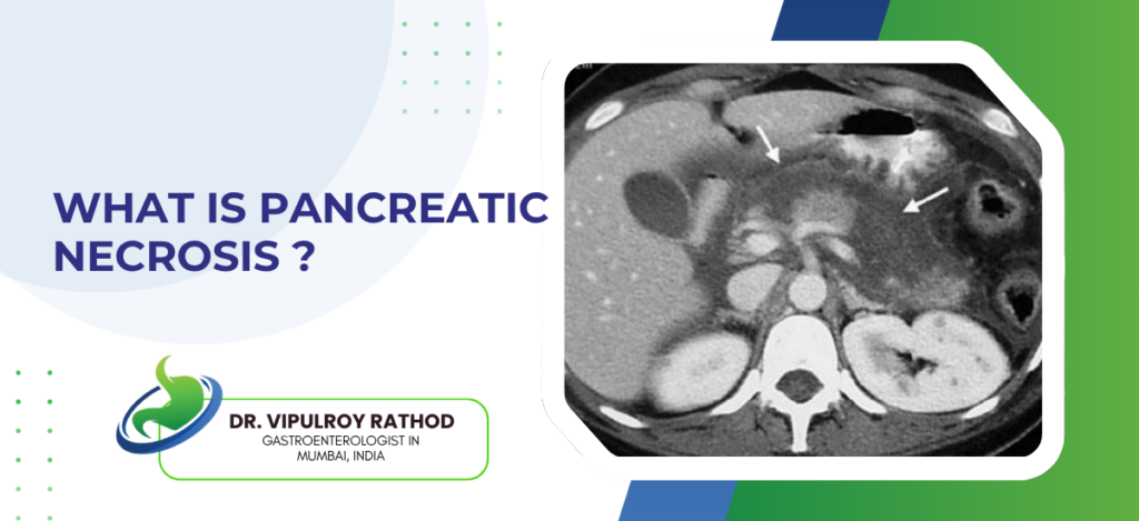
Pancreatic necrosis, or necrotizing pancreatitis, is increasingly treated without open surgery using a “step-up approach” involving minimally invasive techniques. Key non-surgical methods include endoscopic drainage (using an endoscope to drain fluid into the stomach), percutaneous catheter drainage (using imaging to place drains through the skin), antibiotics for infection, and intensive supportive care (fluid resuscitation and early enteral nutrition). According to Dr. Vipulroy Rathod, an experienced Gastroenterologist in Mumbai, “Open surgery for pancreatic necrosis used to be the only option and it carried serious risks, but with endoscopic necrosectomy we can now clear dead tissue through a scope with far better outcomes for the patient.” What endoscopic treatments are used for pancreatic necrosis? Picking the right technique comes down to where the collection sits, how old it is, and whether infection has set in. EUS-guided drainage: An endoscope fitted with ultrasound finds the collection from inside the stomach. A stent gets placed across the stomach wall into the pocket of necrotic fluid. Everything drains gradually over days. No skin incision, no operating theatre involved. Direct endoscopic necrosectomy: After the drainage stent is working, solid necrotic debris still has to come out. The scope goes right through the stent tract into the cavity and scoops out dead tissue. It rarely finishes in one session. Two, three, sometimes four sessions over weeks is normal. Lumen-apposing metal stents: These are a newer design made for exactly this job. The opening is wider than plastic stents so the scope can pass through easily. They hold the tract open long enough for multiple clean-out sessions without needing repeated stent changes. Percutaneous and endoscopic combined: Sometimes the collection stretches into spaces the scope physically cannot reach. In those situations a radiologist places a drain through the skin for the extra pockets while the endoscopist handles the rest. Both teams work together rather than choosing one over the other. Patients dealing with necrotising pancreatitis really do need specialist input, and the pancreatitis treatment page covers how conservative and endoscopic options are matched to each individual case. Why is non-surgical treatment preferred now? Open surgery for pancreatic necrosis carried very high mortality in older data. The endoscopic approach changed that and the numbers behind the shift are strong enough that most specialist centres now go non-surgical by default. Lower mortality: Multiple randomised trials now show endoscopic necrosectomy gives lower mortality than open surgical necrosectomy. That alone moved practice in most tertiary centres. More recent data has only reinforced the trend. Fewer complications afterwards: Less new-onset organ failure. Less bleeding in the post-op period. Fewer secondary infections. ICU stays end up noticeably shorter than what surgery needs for the same diagnosis. Quicker recovery, shorter hospital stay: No abdominal wound means patients are mobilising within days, not weeks. Discharge happens earlier. Functional recovery at 3 and 6 months reads better in follow-up data too. Healthy pancreas gets preserved: Open surgical necrosectomy often ends up damaging viable pancreas while removing the dead parts. Endoscopic tools touch only what’s dead. The difference matters because patients losing functional pancreas develop diabetes or malabsorption later. This move away from open surgery isn’t unique to the pancreas, and the achalasia and POEM blog goes into another condition where endoscopy replaced what used to be a major open operation. Why choose Dr. Vipulroy Rathod for pancreatic necrosis treatment? Dr. Vipulroy Rathod has over 20,000 EUS procedures behind him across 30 plus years of practice. Necrotising pancreatitis is exactly the category of case that benefits most from that depth, because the timing and sequencing of each intervention is what determines how well a patient ends up doing. The experience goes beyond the technical work. Families get the situation explained properly. Decisions are discussed before being made. What recovery actually looks like week by week is laid out honestly. Book your consultation today with one of India’s most experienced pancreatic endoscopy specialists. Book Appointment Call now Frequently Asked Questions Can pancreatic necrosis always be treated without surgery? Most cases can be managed endoscopically now, but complex situations with extensive necrosis may still require combined or surgical approaches depending on clinical factors. How long does endoscopic necrosectomy take to complete? Full treatment usually requires multiple sessions spread over several weeks, though many patients begin feeling significantly better after the first drainage procedure itself. Is endoscopic drainage painful? The procedure is performed under sedation so there’s no pain during it, and most patients experience only mild discomfort for a day or two afterwards. What is the success rate of endoscopic necrosectomy? Success rates in experienced centres consistently cross 80 to 90 percent depending on patient selection and how promptly the treatment is initiated. Reference links- Management of Acute Necrotizing Pancreatitis — American College of Gastroenterology Guidelines Endoscopic Necrosectomy Clinical Evidence — National Library of Medicine

How Is Pancreatic Necrosis Treated Without Surgery Read More »

Who Needs Colon Cancer Screening in India

Colon cancer screening in India is recommended for all adults starting at age 45, even without symptoms. People with a family history of colon cancer, inflammatory bowel disease, or genetic syndromes should start earlier. Anyone with persistent changes in bowel habits, rectal bleeding, or unexplained weight loss should be evaluated regardless of age. According to Dr. Vipulroy Rathod, an experienced Gastroenterologist in Mumbai, “Colon cancer in India is being diagnosed at younger ages and at more advanced stages than it should be, which is why screening isn’t just a Western guideline to follow but a real need we see walking into our clinic every week.” https://www.youtube.com/shorts/0jImZw3wNIw Who should get screened at the standard age? The 45 cutoff applies to most Indian adults at average risk though plenty of people in this group still put off booking a screening even when they clearly fit the criteria. Average-risk adults over 45: If you’ve got no family history of colon cancer and no IBD and no prior polyps along with nothing off about your bowels, you still fall under standard screening once 45 arrives since polyps can sit quietly inside a perfectly healthy body without ever flagging themselves. Men and women equally: Screening applies to both sexes without much difference in India given that colon cancer figures don’t tilt heavily toward men the way some other cancers do, so any woman thinking she’s automatically lower-risk is working off a misconception rather than actual data. Urban and rural residents: Screening eligibility has spread beyond big-city populations over the last decade or so as food habits across tier-2 cities and smaller towns have shifted toward patterns that mirror urban risk factors, meaning people outside the metros can no longer assume they’re in the safer bucket. Adults with healthy lifestyles: A clean diet and regular workouts lower your statistical risk on paper but won’t take it down to zero, so even the most active person past 45 benefits from having that baseline scope done once just to confirm nothing unexpected is brewing. If you’re over 45 and haven’t scheduled one, a colonoscopy is the most direct way of finding out whether things inside match how healthy everything appears from the surface. Who needs earlier or more frequent screening? A sizeable chunk of the patients coming into gastroenterology clinics across India aren’t average-risk at all with their screening needing to begin much earlier than the standard 45 timeline. Family history of colon cancer: Screening starts 10 years before your parent or sibling’s diagnosis age or by 40 itself whichever comes first, and specialists consider this a firm clinical recommendation rather than something you’re supposed to debate since first-degree family history genuinely alters the risk profile in ways blood tests won’t catch. Inflammatory bowel disease patients: Once Crohn’s or ulcerative colitis has been active in the colon for 8 years or longer cancer risk climbs to a level where surveillance colonoscopies every 1 to 3 years get built into regular disease management rather than standing as a separate screening you schedule alongside everything else. Genetic syndromes like Lynch or FAP: Inherited conditions such as Lynch syndrome and familial adenomatous polyposis reset the screening age entirely with scopes often starting in the teens or early twenties at intervals much tighter than anything that applies to the average Indian adult. Metabolic risk factors: Obesity paired with type 2 diabetes and fatty liver disease keeps showing up in newer studies as connected to higher colon cancer risk which carries real weight for anyone already being treated for two or more of these conditions simultaneously. If you match any of these profiles don’t sit on the screening conversation, and our blog on fatty liver and diabetes is worth a read since it walks through how overlapping metabolic issues can quietly pile up into bigger long-term risks including various cancers. Why choose Dr. Vipulroy Rathod for colon cancer screening? Dr. Vipulroy Rathod has been in gastroenterology for over thirty years with more than 80,000 endoscopic procedures done and thousands of those being colonoscopies performed at Mumbai’s leading hospitals, which translates into the kind of reading of subtle findings that genuinely matters when a small polyp or unusual patch shows up during an otherwise routine screening scope. What patients mention most often isn’t really about the scope itself but about the after-part where results actually get explained properly, the follow-up plan is laid out without vague medical talk, and concerns get real answers rather than the rushed response most people are used to getting in a busy clinic. Book your colon cancer screening today with one of India’s most experienced gastroenterologists. Book Appointment Call now Frequently Asked Questions At what age should colon cancer screening start in India? Most adults should start at 45, but if you have family history or genetic risk factors your doctor may recommend starting earlier than that. Is colon cancer common in India? Colon cancer rates in India have been rising steadily over the past two decades, especially in urban populations with changing dietary and lifestyle patterns.   How often is screening needed? Normal colonoscopy results usually mean one screening every 10 years, but higher-risk patients might need rescreening every 1 to 5 years depending on findings. Is colon cancer screening covered by insurance in India? Most private health insurance policies cover colonoscopy when recommended by a doctor, though coverage varies and it’s worth confirming specifics with your insurer beforehand. Reference links- Colorectal Cancer Screening Recommendation — U.S. Preventive Services Task Force (USPSTF) Colorectal Cancer in India: Epidemiology and Screening — World Health Organization Global Cancer Observatory

Who Needs Colon Cancer Screening in India Read More »

Bile Leakage After Gallbladder Surgery: Causes, Symptoms, and Treatment

Gallbladder surgery, also known as cholecystectomy, is a widely performed procedure to treat gallstones, inflammation, and related digestive issues. It is generally safe and helps restore normal digestion by removing the diseased gallbladder. However, like any surgical procedure, certain complications may occur during the healing phase. One such complication is bile leakage after gallbladder surgery, where bile escapes from the bile ducts into the abdominal cavity instead of flowing into the intestine. This can lead to pain, infection, and delayed recovery if not addressed promptly. Dr. Vipulroy Rathod,a highly respected gastroenterologist in Mumbai, India, highlights, “Early identification of bile leakage is essential to prevent complications and ensure smooth healing.” He further adds, “With advanced endoscopic techniques, most cases can be managed effectively without the need for major surgery.” Dr. Rathod is widely recognized for his precision in managing complex biliary and pancreatic conditions. With extensive experience in advanced endoscopic procedures such as ERCP and EUS, he focuses on resolving post-surgical complications with minimally invasive techniques. His approach to managing bile leakage after gallbladder surgery emphasizes early intervention, accurate diagnosis, and targeted treatment, helping patients recover efficiently while minimizing the need for repeat procedures or prolonged hospitalization. Why does bile leakage happen after gallbladder removal? Let’s explore the common reasons behind this condition. What Causes Bile Leakage After Gallbladder Surgery? Bile leakage typically occurs when there is disruption or injury to the bile ducts during or after cholecystectomy. While it is not very common, certain factors can increase the risk. Common causes include:        Injury to bile ducts:Accidental damage during surgery can lead to leakage.        Cystic duct stump leak:Improper sealing of the duct after gallbladder removal may cause bile to escape.        Accessory bile ducts:Small, unnoticed ducts can leak bile if not identified during surgery.        Inflammation or infection:Pre-existing inflammation can weaken duct structures.        Surgical complications:Complex procedures may increase the risk of bile duct disruption. Understanding these causes helps in early detection and timely bile leakage treatment. Experiencing discomfort after surgery? Connect with a specialist to determine the underlying cause and next steps. Book Appointment Call now What signs should you watch for after surgery? Let’s discuss the symptoms that may indicate bile leakage. Common Symptoms of Bile Leakage After Gallbladder Surgery Symptoms of bile leakage can vary depending on the severity, but early warning signs should never be ignored. Common symptoms include:        Persistent abdominal pain, especially in the upper abdomen        Fever or chills, indicating possible infection        Nausea and vomiting        Swelling or bloating in the abdomen        Jaundice (yellowing of skin and eyes)        Drainage of bile fluid from surgical wounds (in some cases) “Recognizing these symptoms early can prevent complications and ensure faster recovery,” states Dr. Vipulroy Rathod. Now, let’s discuss the typical timeline of bile leakage after surgery. When Does Bile Leakage Typically Occur After Surgery? Bile leakage usually develops within the first few days after cholecystectomy, but in some cases, symptoms may appear later. Typical timeline:        Within 2–5 days:Most common period for symptoms to begin        Within 1–2 weeks:Delayed symptoms may occur in some patients        Rare cases:Late presentation if leakage is mild and gradual Early follow-up after surgery is important to detect any complications during this critical period. Let’s explore the diagnostic methods used to identify this condition. How is Bile Leakage Diagnosed? Accurate diagnosis is essential for effective bile leakage treatment and involves a combination of clinical evaluation and imaging. Diagnostic methods include: Ultrasound:Helps detect fluid accumulation in the abdomen CT scan:Provides detailed imaging of bile leakage and surrounding structures HIDA scan:Tracks bile flow to identify leaks MRCP (MRI scan):Visualizes bile ducts without invasive procedures ERCP (Endoscopic Retrograde Cholangiopancreatography):Both diagnostic and therapeutic These tests help locate the leak and guide appropriate treatment. How is bile leakage treated effectively? Let’s explore the treatment approaches available. Treatment Options for Bile Leakage After Gallbladder Surgery Treatment depends on the severity and source of the leak, with most cases managed using minimally invasive techniques. The treatment options include: Endoscopic Treatment (ERCP) This is the most commonly used approach, where an endoscope is used to access the bile duct and place a stent to redirect bile flow and reduce leakage. Biliary Stenting A small tube is inserted into the bile duct to keep it open and allow bile to flow correctly into the intestine, helping the leak to heal naturally. Sphincterotomy A minor procedure is done during ERCP in which a small cut is made to ease bile flow and reduce the pressure within the bile ducts. Percutaneous Drainage In case bile has built up in the abdomen, a drain may be placed through the skin to clear the fluid and prevent infection. Antibiotic Therapy Antibiotics are prescribed to treat or prevent infections caused by bile leakage. Surgical Repair (Rare Cases) Surgery can be performed to repair the bile duct in critical or complicated cases where minimally invasive intervention is not effective. Supportive Care and Monitoring Regular checkups, imaging and monitoring will be done to make sure that the leak is healing well and no further complications develop. Early and appropriate bile leakage treatment significantly improves recovery and reduces the risk of complications. What happens if bile leakage is ignored? Let’s understand the potential risks Complications if Bile Leakage is Left Untreated Untreated bile leakage can lead to serious complications, making early treatment essential. Possible complications include: Infection or abscess formation Peritonitis (inflammation of abdominal lining) Sepsis, a severe systemic infection Persistent abdominal pain and discomfort Delayed recovery and prolonged hospitalization Timely intervention can prevent these complications and improve outcomes. Worried about complications? Get in touch with a professional to avoid serious health risks and ensure smooth healing and long-term wellness. Book Appointment Call now What does recovery look like after treatment? Let’s explore the healing process Recovery and Healing After Bile Leak Treatment Recovery after bile leakage treatment depends on the severity of the condition and the type of treatment performed. Recovery highlights: Most patients recover well with minimally invasive procedures Symptoms improve within a few days after treatment Regular follow-up ensures proper healing Temporary stents may be removed

Bile Leakage After Gallbladder Surgery: Causes, Symptoms, and Treatment Read More »

What Is Achalasia And Why Does POEM Work

Achalasia is a rare oesophageal motility disorder where the lower oesophageal sphincter fails to relax during swallowing and the oesophagus loses its coordinated muscle contractions, causing progressive difficulty swallowing both solids and liquids, regurgitation of undigested food, chest pain, and significant weight loss over time, POEM (Per-Oral Endoscopic Myotomy) works because it cuts the dysfunctional sphincter muscle from inside the oesophageal wall without any external incision, directly resolving the obstruction that medication and dilation can only temporarily manage. According to Dr. Vipulroy Rathod, Gastroenterologist in Mumbai, “Achalasia is one of those conditions where patients spend years being treated for reflux or anxiety before anyone thinks to investigate the oesophagus properly, and POEM changed the treatment landscape because it addresses the actual mechanical problem rather than working around it.” What Is Achalasia and How Does It Develop? Damaged nerve cells in the oesophageal wall are the root cause and once those cells are gone they don’t regenerate, which is why achalasia is managed not cured and why the intervention needs to be durable rather than temporary. Mechanism: Myenteric plexus neurons that coordinate lower oesophageal sphincter relaxation are progressively destroyed, likely through autoimmune processes in most cases, leaving a sphincter that stays contracted during swallowing and an oesophagus that can’t move food downward through organised peristalsis. Symptoms: Progressive dysphagia for both solids and liquids is the hallmark, regurgitation of undigested food hours after eating, chest pain from oesophageal spasm, and weight loss that gets attributed to anxiety or functional disorders for months before anyone orders a proper motility study. Diagnosis: High-resolution manometry is the gold standard showing absent peristalsis and incomplete LES relaxation, barium swallow shows the classic bird-beak narrowing at the gastro-oesophageal junction, endoscopy rules out malignancy causing pseudoachalasia which has identical symptoms but a completely different clinical picture. Types: Chicago Classification divides achalasia into three types based on manometry pattern, Type II with pan-oesophageal pressurisation responds best to POEM, Type I and III also respond well, and knowing which type the patient has changes how the myotomy length and approach gets planned by the endoscopist. Achalasia diagnosis changes what treatment options exist and which ones are actually appropriate for that patient’s specific manometry pattern. Specialist in endoscopy investigates and stages properly before any intervention gets planned rather than defaulting to dilation because it’s the simpler first step. Why Does POEM Work Better Than Other Treatments? Dilation and Botox buy time. POEM fixes the mechanical problem and the outcomes reflect that difference consistently across the literature. Myotomy: POEM creates a submucosal tunnel inside the oesophageal wall, cuts the circular muscle fibres of the lower oesophageal sphincter under direct endoscopic vision, and the sphincter that was keeping food out relaxes permanently without any external incision or surgical access needed. Durability: Long-term success rates above 85 to 90% at five years in most published series, significantly better than pneumatic dilation which needs repeat sessions in a substantial proportion of patients and Botox injection which typically wears off within months and needs repeating indefinitely. Recovery: No external incision means no wound, patients typically eat the following day, discharged within 48 hours in most cases, and the recovery profile looks nothing like surgical Heller myotomy which achieves similar results but requires laparoscopic access and a longer recovery. Reflux: POEM does carry higher post-procedure reflux rates than Heller myotomy with fundoplication because no anti-reflux procedure accompanies it, and patients need to understand this going in and commit to post-procedure pH monitoring and proton pump inhibitor management rather than treating POEM as a complete solution with no follow-up required. POEM is the most effective single intervention for achalasia and patients who understand it commit to post-procedure care far better. Similar endoscopic precision drives neurolysis for pancreatic pain without any incision. Why Choose Dr. Vipulroy Rathod Dr. Vipulroy Rathod has been performing advanced therapeutic endoscopy including third space endoscopy procedures at Fortis Hospital Mulund for over 30 years, with the EUS and endoscopic expertise that complex procedures like POEM require in terms of submucosal dissection technique, real-time decision-making during the tunnel creation, and managing any complication that arises without converting to open surgery, trained physicians from 35 countries in exactly this. Patients arrive having been managed with dilation for years with diminishing returns and most leave with a procedure that addressed the mechanical problem directly rather than working around it indefinitely. Start Your Treatment Journey Today Book Appointment Call now Frequently Asked Questions What are the first symptoms of achalasia? Progressive difficulty swallowing both solids and liquids, regurgitation of undigested food, and chest pain are the most common early symptoms. How is achalasia diagnosed? High-resolution manometry is the gold standard, supported by barium swallow showing bird-beak narrowing and endoscopy to exclude malignancy. Is POEM permanent for achalasia? POEM achieves durable relief in over 85% of patients at five years making it the most effective long-term treatment currently available for achalasia. Does POEM cause acid reflux? POEM increases reflux risk compared to surgical myotomy with fundoplication and patients need ongoing proton pump inhibitor therapy and pH monitoring after the procedure. Reference links- Achalasia Diagnosis and Management — American Society for Gastrointestinal Endoscopy POEM Procedure Guidelines — World Gastroenterology Organisation

What Is Achalasia And Why Does POEM Work Read More »

Is Fatty Liver Linked to Diabetes

Yes, fatty liver and diabetes are directly linked through insulin resistance, the same metabolic dysfunction that drives fat accumulation in the liver also impairs glucose regulation, both conditions develop together and each makes the other worse, Type 2 diabetics have significantly higher rates of MASLD than the general population and people with fatty liver carry elevated diabetes risk even when their blood sugar currently looks fine on a report. According to Dr. Vipulroy Rathod, Gastroenterologist in Mumbai, “Fatty liver and diabetes are two expressions of the same underlying metabolic problem and treating one without addressing the other is why so many patients cycle through management plans that control numbers on paper without actually changing what’s happening in the liver.” https://www.youtube.com/shorts/i2roa-d-Ojs How Are Fatty Liver and Diabetes Connected? Same root cause, two organs expressing it differently, and most patients don’t find out the connection exists until both have been managed separately for years without either improving properly. Resistance: Liver cells stop responding to insulin, accumulate fat, the pancreas compensates by producing more which drives further fat storage, and that loop runs quietly until blood sugar can no longer be maintained regardless of how much insulin gets produced. Bidirectional: Fatty liver drives insulin resistance which drives diabetes, but existing diabetes accelerates liver fat accumulation and fibrosis progression simultaneously, so both get worse together unless both get properly managed together and most treatment plans don’t do that. MASH: Type 2 diabetics with fatty liver are far more likely to progress to metabolic steatohepatitis than fatty liver patients without diabetes, and most of them have no idea their diabetes is actively changing their liver disease trajectory while they’re managing blood sugar as a standalone problem. Fat: Central obesity deposits fat around internal organs including the liver, drives the inflammatory signals that damage liver tissue and impair beta cell function at the same time, and dietary advice given without structured metabolic intervention rarely reverses this in practice regardless of how motivated the patient is at the start. Managing fatty liver without looking at the metabolic picture underneath is treating a symptom not a disease. Specialist in fatty liver assesses the full metabolic context rather than just noting what the scan shows and stopping there. How Should Both Conditions Be Managed Together? Treating this as one metabolic problem rather than two separate referrals is what changes what management actually achieves for these patients. Weight: 5 to 10% body weight reduction improves liver fat, reduces fibrosis risk, and improves insulin sensitivity at the same time, and this is why structured weight loss works where generic lifestyle advice given at the end of a consultation consistently doesn’t. Medication: GLP-1 receptor agonists improve blood sugar, drive meaningful weight loss, and reduce liver fat through one mechanism rather than requiring separate drug regimens for two conditions that most patients can’t realistically maintain long term anyway. Monitoring: Both conditions together means fibrosis staging through fibroscan or FIB-4 blood markers, not just a liver ultrasound that tells the patient fat is present without telling them where on the fibrosis spectrum they actually sit or how urgently that matters. Alcohol: Even moderate drinking in a patient with both fatty liver and diabetes accelerates liver damage significantly beyond what either condition does alone, and most patients underestimate this because nobody tells them directly that their threshold is much lower than someone without their metabolic profile. Both conditions share the same root and addressing that root changes the trajectory of both. Read more on MASLD to understand the full picture of metabolic fatty liver disease and what proper management actually looks like. Why Choose Dr. Vipulroy Rathod Dr. Vipulroy Rathod has been managing fatty liver and its metabolic connections including diabetes and insulin resistance for over 30 years at Fortis Hospital Mulund, staging liver disease through fibroscan and blood markers rather than stopping at the ultrasound, building management plans that address the metabolic drivers rather than monitoring the liver while the underlying problem continues, trained physicians from 35 countries in this. Patients arrive having been told separately that they have fatty liver and that they have diabetes and most leave understanding for the first time that these are two expressions of one problem with one plan that actually addresses both. Start Your Treatment Journey Today Book Appointment Call now Frequently Asked Questions Does diabetes cause fatty liver? Yes, insulin resistance in Type 2 diabetes drives fat accumulation in the liver and significantly increases MASLD risk and progression rate. Can treating fatty liver improve diabetes control? Yes, reducing liver fat through weight loss and medication improves insulin sensitivity and often leads to better blood sugar control in Type 2 diabetics. How common is fatty liver in diabetic patients? Around 50 to 75% of Type 2 diabetic patients have some degree of fatty liver making it one of the most common complications of diabetes. Should diabetic patients get liver fibrosis testing? Yes, diabetic patients with fatty liver should have fibrosis staging through fibroscan or FIB-4 blood markers not just ultrasound given their higher progression risk. Reference links- Fatty Liver and Diabetes Connection — American Association for the Study of Liver Diseases MASLD and Metabolic Disease Guidelines — European Association for the Study of the Liver

Is Fatty Liver Linked to Diabetes Read More »

Scroll to Top
Call Now Button