Dr. Vipulroy Rathod

Author name: Dr. Rathod Medical Foundation

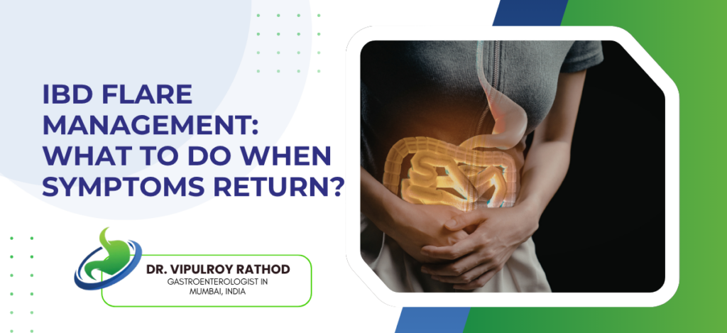
IBD Flare Management: What to Do When Symptoms Return?

A flare is the return of active IBD symptoms after a period of remission. Patients usually know what their flare looks like, because the symptoms tend to follow the same pattern that brought them to a doctor in the first place. Stool frequency rises, blood may reappear, abdominal pain creeps back, fatigue and weight loss follow if the flare runs long enough. The instinct to wait it out is one of the commoner mistakes patients make. Acting early shortens flares and prevents the damage that’s much harder to undo later. According to Dr. Vipulroy Rathod, Gastroenterologist in Mumbai, “Patients wait too long. They hope it’ll settle on its own. Sometimes it does. Often it doesn’t, and what could have been managed with a steroid course or a biologic dose tweak in week one ends up needing hospitalisation by week four. Acting early when it turns out to be nothing costs much less than acting late when it turns out to be a major flare.” What to do when symptoms return? The first response isn’t a Google search. It’s a structured assessment, ideally with input from the gastroenterology team within days, not weeks. Document symptoms specifically. Stool frequency per day. Presence and amount of blood. Pain pattern. Weight change. Vague reports like “things are worse” don’t give the clinical team enough to work with. Check medication adherence honestly. Missed doses, stopped biologics, irregular maintenance therapy, these are the commonest preventable causes of flare. Patients are often reluctant to admit gaps, but the team needs to know because management changes depending on whether the flare comes from missed treatment or genuine breakthrough disease. Always rule out infection before escalating immunosuppression. Stool testing for C. difficile, bacterial pathogens, and parasites is non-negotiable before adding steroids. Giving steroids to a patient with superimposed C. difficile colitis makes things significantly worse. Contact the gastroenterology team early. Many flares can be assessed remotely at first, with stool tests, blood markers, and history guiding the next step. IBD treatment plans should include clear flare instructions agreed in advance, telling patients which symptoms warrant immediate contact, what tests will be ordered, and what the escalation threshold looks like, so the response is structured rather than reactive. What clinical assessment and escalation actually involves? Once a flare is reported, the workup follows a familiar pattern. Confirm active inflammation. Rule out other causes. Decide how aggressively to escalate. Stool tests come first because they answer the most important question quickly. Faecal calprotectin to quantify inflammation. Cultures and PCR for infection. Calprotectin above 250 in a patient with returned symptoms strongly suggests active disease. Under 100 raises the possibility that something else is going on entirely, sometimes IBS-like symptoms in patients with healed bowel, sometimes a non-IBD cause that’s been missed. Blood tests cover inflammatory markers (CRP and ESR), full blood count, liver function, and a basic metabolic panel. Anaemia, low albumin, raised CRP all support active disease. In biologic patients drug levels go in at the same time, because the commonest reason for biologic failure isn’t drug resistance, it’s underdosing. Imaging or endoscopy isn’t always needed at first contact. Mild to moderate flares are often managed on stool and blood tests alone in the early phase. Severe symptoms, suspected complications, or flares not responding to first-line treatment usually trigger CT enterography, MR enterography, or sigmoidoscopy depending on availability and what’s clinically most useful. Treatment escalation runs on a ladder rather than a single decision. Optimise maintenance therapy. Add a short course of steroids if needed for symptom control. Consider biologic dose escalation or switching agents if the flare reflects genuine loss of response. Surgery for cases that don’t respond to medical rescue, or where complications have developed. Read more on when IBD needs surgery for how the surgical option fits into flare management when medical treatment isn’t enough. Why choose Dr. Vipulroy Rathod for IBD flare management? Dr. Vipulroy Rathod has been managing IBD flares and the longer trajectory of inflammatory bowel disease at Fortis Hospital Mulund for over three decades, with the kind of structured flare protocols that come from seeing the same patterns repeatedly and knowing which escalation works for which patient. Many patients arrive mid-flare having waited too long, when earlier intervention would have made the difference between outpatient management and a hospital admission. The pattern that matters most in flare management is recognition. Patients who know what their flare looks like, who have a clear plan agreed with their team, who contact the clinic at day three rather than week three, consistently do better than patients trying to manage flares alone. The work upfront on agreeing what to do when symptoms return pays back many times over. Book your consultation today with one of India’s most experienced specialists for IBD assessment and structured flare management. Book Appointment Call now Frequently Asked Questions How quickly should I contact the clinic during a flare? Within a few days. Don’t wait for symptoms to become severe. Early assessment gives the team more options than late assessment. Can I just take more of my current medication? Sometimes, often not. Some flares respond to maintenance optimisation. Others need short steroid courses or biologic adjustment. Self-adjusting without team input risks under-treating or masking infection. What blood and stool tests are usually done? Faecal calprotectin and stool infection screen first. Then CRP, full blood count, liver function. Biologic drug levels if applicable. Most results come back within a few days. Will I need hospital admission? Most flares are managed as outpatients. Admission is reserved for severe colitis not responding to oral treatment, complications like obstruction or perforation, or patients too unwell to manage at home. Reference links- IBD Flare Management Guidelines, American Gastroenterological Association — https://gastro.org/clinical-guidance/ ECCO Topical Review on IBD Flare, European Crohn’s and Colitis Organisation — https://www.ecco-ibd.eu/publications/ecco-guidelines.html

IBD Flare Management: What to Do When Symptoms Return? Read More »
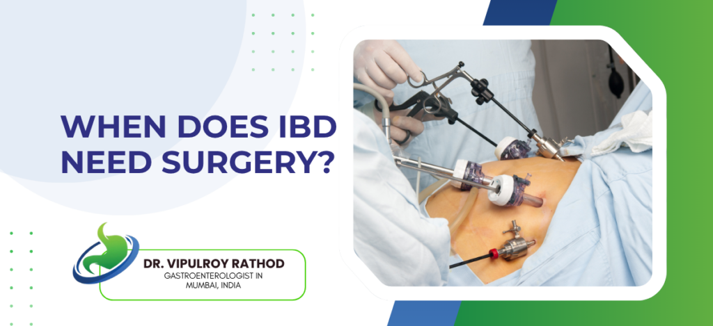
When Does IBD Need Surgery?

  Surgery in inflammatory bowel disease isn’t a failure of medical treatment. It’s a tool used when medical therapy can’t control the disease, when complications develop, or when the damage already done has become structural. Around twenty to thirty percent of ulcerative colitis patients eventually need a colectomy, and the figure rises to fifty percent or more in Crohn’s disease over a lifetime. The decision is rarely simple. It depends on disease pattern, response to medication, the surgeon’s view, and what the patient is willing to live with. According to Dr. Vipulroy Rathod, Gastroenterologist in Mumbai, “The right time for IBD surgery is a conversation, not a calculation. Some patients are pushed into surgery too early because someone gave up on medication too quickly. Others are kept on failing biologics for years when surgery would have given them their life back. Getting that timing right depends on knowing both the medical and the surgical sides equally well.” When surgery becomes necessary in IBD? The indications fall into urgent and elective categories, and the decision-making works differently for each. Acute severe colitis not responding to medical rescue. A patient hospitalised with severe ulcerative colitis who doesn’t respond to intravenous steroids or infliximab rescue within three to five days needs urgent colectomy. Continuing medical therapy beyond that window risks colonic perforation, toxic megacolon, or death. Bowel obstruction from Crohn’s strictures is the commonest reason for elective surgery in Crohn’s disease. Long-standing inflammation leaves fibrotic narrowings that don’t respond to medication, and the patient develops recurrent obstructive symptoms that medical treatment can’t relieve. Resection or strictureplasty becomes the answer. Perforation, abscess, or fistula complications. A perforated bowel from severe IBD needs emergency surgery without delay. Localised abscesses can sometimes be drained percutaneously first, with planned resection later, but the underlying anatomy usually still needs surgical correction. Dysplasia or cancer on surveillance colonoscopy. Long-standing ulcerative colitis raises colorectal cancer risk significantly, and high-grade dysplasia or invasive cancer found at surveillance means colectomy regardless of how the disease is currently behaving. The challenge is that these decisions often look obvious in retrospect but aren’t always obvious in the moment. IBD treatment planning that integrates the gastroenterologist’s medical view with the IBD surgeon’s anatomical view tends to produce better timing decisions than either specialty working in isolation. What surgery actually looks like for different IBD patients? The type of surgery depends on which IBD it is and what’s being treated, and the outcomes vary considerably between procedures. For ulcerative colitis, surgery means removing the colon entirely because the disease only affects the colon and rectum. The standard procedure is a restorative proctocolectomy with ileal pouch-anal anastomosis (IPAA), creating a J-pouch from the small bowel that connects to the anus and allows the patient to pass stool normally without a permanent stoma. Most patients adapt well over six to twelve months, with reasonable continence and quality of life thereafter. Crohn’s disease surgery works very differently because the disease can affect any part of the gut and recurs in around fifty percent of patients within five years even after successful resection. The surgical principle is to remove as little as possible. Limited resection of the diseased segment, often the terminal ileum, or strictureplasty where the bowel is widened without being removed. Aggressive surgery to “cure” Crohn’s doesn’t work and produces short-bowel problems that are often worse than the disease. There are situations where a permanent stoma is the right answer. Severely damaged rectum that can’t be reconnected. Recurrent pouch failure in ulcerative colitis. Perianal Crohn’s that destroys sphincter function. For these patients, a permanent ileostomy isn’t a defeat. It’s often the operation that gives them their quality of life back after years of fighting the disease. The conversation about surgery should include the surgeon early, not as a last resort. Read more on biologics for IBD for how modern medical therapy fits alongside surgical options in the overall treatment plan. Why choose Dr. Vipulroy Rathod for IBD surgical decision-making? Dr. Vipulroy Rathod has been managing IBD patients through medical therapy and the decisions around surgery at Fortis Hospital Mulund for over three decades, with the coordinated approach between gastroenterology and IBD surgery that this disease genuinely requires. Many patients arrive having been kept on failing medications for too long, or pushed towards surgery before the medical options were fully tried, depending on which specialty saw them first. The decision about IBD surgery isn’t a binary one. It’s a judgment about timing, disease behaviour, patient preference, and what the future trajectory looks like with and without an operation. The hardest part is often acknowledging that medical therapy has done what it can, which most gastroenterologists are reluctant to admit too early, and most surgeons too willing to declare too readily. Book your consultation today with one of India’s most experienced specialists for IBD assessment and integrated medical-surgical management. Book Appointment Call now Frequently Asked Questions Does every IBD patient eventually need surgery?  No. Many never do. Around fifty to seventy percent of ulcerative colitis patients and fifty percent of Crohn’s patients avoid surgery entirely over their lifetime, particularly with modern biologic therapy. Is IBD surgery a cure? For ulcerative colitis, yes, in the sense that removing the colon removes the disease. For Crohn’s disease no, the disease recurs in around half of patients within five years even after successful resection. Can IBD surgery be avoided once it’s been recommended? Sometimes, particularly in elective situations where there’s room for further medical optimisation. In urgent situations like perforation or severe colitis not responding to rescue therapy, surgery can’t safely be delayed. How long is recovery after IBD surgery? Hospital stay is usually five to ten days. Return to normal activity around four to six weeks for straightforward resections, longer for pouch surgery or staged procedures. Adaptation to a pouch or stoma can take six to twelve months for full adjustment. Reference links- IBD Surgery Guidelines, American Society of Colon and Rectal Surgeons — https://fascrs.org/healthcare-providers/clinical-practice-guidelines ECCO Guidelines on Surgical Management, European

When Does IBD Need Surgery? Read More »

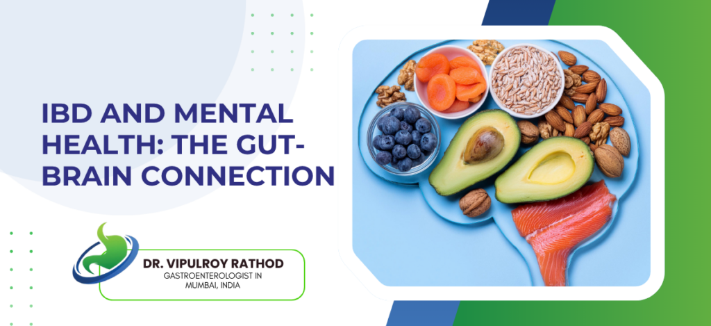
IBD and Mental Health: The Gut-Brain Connection

Inflammatory Bowel Disease (IBD)—including Crohn’s disease and ulcerative colitis—is deeply linked to mental health through the gut-brain axis. Chronic intestinal inflammation disrupts this two-way communication system, significantly increasing the risk of anxiety and depression, while psychological stress can actively trigger or worsen physical IBD flare-ups. According to Dr. Vipulroy Rathod, Gastroenterologist in Mumbai, “I’ve seen patients whose IBD only stabilises once their anxiety or depression gets properly treated. And the reverse, patients whose mood improves substantially when their gut inflammation finally comes under control. The gut-brain connection isn’t theoretical anymore. Ignoring the mental health side is one of the commoner reasons patients don’t respond to treatment the way they should.” How the gut and brain talk to each other in IBD? The gut-brain axis is a real anatomical and biochemical system, not a metaphor. Several pathways connect the two. Inflammatory cytokines crossing the blood-brain barrier. TNF-alpha, interleukin-6, and similar molecules produced during active IBD reach the brain and directly affect mood-regulating circuits. This is part of why flares often come with worsening fatigue and low mood before the bowel symptoms themselves become severe. The vagus nerve carries signals in both directions between the gut and the central nervous system. Pain perception, satiety, emotional regulation, autonomic function. In active IBD this signalling is disrupted, and many of the symptoms patients attribute to the disease itself, fatigue, poor sleep, altered stress tolerance, come partly from this disruption. Gut microbes and neurotransmitters. Most of the body’s serotonin is made in the gut. So is a meaningful proportion of GABA precursors. Dysbiosis in IBD shifts the production of these chemicals, which is one biological link between microbiome changes and mood symptoms that wasn’t recognised for years. HPA axis dysregulation. Chronic IBD activates the cortisol stress response continuously, and that chronic activation contributes to anxiety, sleep problems, and reduced capacity to handle stress, which then feeds back into disease activity. Understanding the mechanism changes treatment priorities. IBD treatment plans that include mood screening at every clinic visit, with appropriate referral or treatment when something is identified, produce meaningfully better outcomes than gastroenterology-only care. What this means for patient care in practice? The clinical consequences show up in how IBD should be managed day to day. Mood screening should happen at diagnosis and at every flare. PHQ-9 for depression, GAD-7 for anxiety, both quick to administer and meaningfully better than asking “how are you feeling” in passing. A surprising proportion of patients screen positive without ever having mentioned mood symptoms, often because they assumed those symptoms were just part of having a chronic disease. Biologic therapy and other anti-inflammatory treatments improve mood substantially in many patients, sometimes more than they improve the gut symptoms. Patients started on infliximab or vedolizumab who report better energy, improved sleep, and reduced anxiety within weeks aren’t experiencing a placebo response. They’re experiencing the central effect of removing inflammatory signalling from the brain. This is real. Psychological treatment, when needed, works. CBT specifically adapted for IBD has good evidence for reducing both anxiety symptoms and disease activity. SSRIs are safe in IBD and combine fine with biologic therapy. The reluctance to start psychiatric medication in IBD patients is mostly outdated thinking that hasn’t caught up with the evidence. Stress management isn’t just lifestyle advice. Mindfulness, regular sleep, exercise, yoga, all have measurable effects on inflammatory markers in IBD, separate from their direct effects on mood. Not substitutes for medication, but not optional extras either. Read more on what IBD is and how it’s diagnosed for how the underlying conditions sit within the broader clinical picture. Why choose Dr. Vipulroy Rathod for IBD and mental health care? Dr. Vipulroy Rathod has been managing inflammatory bowel disease at Fortis Hospital Mulund for over three decades, with the integrated approach to gut and mental health that the modern evidence now demands. Many patients arrive having been treated for years by gastroenterologists who never asked about mood, and separately by psychiatrists who didn’t know much about IBD, with neither side aware that the two were biologically connected. The patients who do best in this disease are usually the ones whose IBD team takes mental health seriously and whose mental health team understands what active gut inflammation does to mood. Not every patient needs a psychiatrist alongside their gastroenterologist, but every patient should be screened, and the ones who need integrated care should get it without delay. Book your consultation today with one of India’s most experienced specialists for IBD management and integrated mental health assessment. Book Appointment Call now Frequently Asked Questions Are anxiety and depression more common in IBD patients? Yes, significantly. Around a third meet criteria for clinical anxiety or depression, compared to ten to fifteen percent in the general population. Does treating mental health improve IBD outcomes? Yes. Better adherence to IBD medication, fewer flares, better quality of life. The evidence is consistent now. Can stress alone trigger an IBD flare? Stress doesn’t cause IBD, but it can trigger flares in established disease. The cortisol and inflammatory signalling pathway is well-mapped at this point. Are antidepressants safe to use with biologics? Yes. SSRIs and most antidepressants combine with biologic therapy without significant interaction concerns. The combination is common in IBD practice. Reference links- IBD and Psychological Comorbidity Position Statement, American Gastroenterological Association — https://gastro.org/clinical-guidance/ Brain-Gut Behavioral Therapies in IBD, Crohn’s and Colitis Foundation — https://www.crohnscolitisfoundation.org/

IBD and Mental Health: The Gut-Brain Connection Read More »

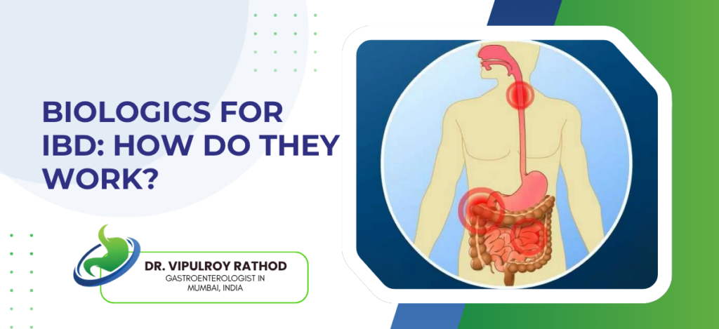
Biologics for IBD: How Do They Work?

Biologics are large protein molecules, usually antibodies, designed to block specific inflammatory pathways that drive Crohn’s disease and ulcerative colitis. Unlike steroids or immunosuppressants which suppress the immune system broadly, biologics target one molecule, one cytokine, one cell type, leaving the rest of the immune system mostly intact. They’ve transformed IBD treatment over the last two decades. Patients who would have ended up with surgery or chronic steroid use now achieve sustained remission on infusions or injections every few weeks. The trade-off is cost and complexity. According to Dr. Vipulroy Rathod, Gastroenterologist in Mumbai, “When biologics work, they really work. Patients on infliximab or vedolizumab who failed years of steroids and immunosuppressants achieve mucosal healing and stay in remission for years. The challenge is matching the right drug to the right patient, and starting it early enough to change the disease course rather than waiting until damage has already accumulated.” How biologics actually work in IBD? The fundamental idea behind biologic therapy is that IBD inflammation is driven by specific molecules in the immune system, and blocking those molecules controls the disease without shutting down everything else. Anti-TNF agents. Infliximab, adalimumab, golimumab. These block tumour necrosis factor alpha, one of the central pro-inflammatory cytokines in IBD. The oldest class of biologics, used since the late 1990s, with the most evidence behind them. Effective in both Crohn’s and ulcerative colitis. Anti-integrin agents work by stopping inflammatory immune cells from entering the gut. Vedolizumab is the main example, gut-selective, with a strong safety profile because it doesn’t affect immune surveillance elsewhere in the body. Particularly useful in patients with infection risks or comorbidities that make broader immunosuppression dangerous. Anti-IL-12/23 and anti-IL-23 agents. Ustekinumab and risankizumab. These block interleukin signalling pathways involved in chronic gut inflammation. Useful in patients who haven’t responded to anti-TNF, with a different side-effect profile and good evidence in both Crohn’s and increasingly in ulcerative colitis. JAK inhibitors are technically small molecules rather than biologics, but they’re often grouped with them because they sit in the same advanced therapy category. Tofacitinib and upadacitinib block intracellular signalling involved in cytokine response. Oral, fast-acting, with their own risk profile that needs careful patient selection. Choosing the right biologic depends on disease subtype, severity, location, and previous treatment response. IBD treatment decisions today usually involve mapping each patient’s disease pattern against the specific biologic profile rather than trying drugs in a fixed sequence. The era of “anti-TNF first, then see what happens” is fading. Who benefits from biologic therapy? Biologics aren’t for every IBD patient, but the threshold for starting them has lowered significantly as evidence accumulates for early aggressive treatment. The clearest indication is moderate-to-severe disease that hasn’t responded adequately to steroids and conventional immunomodulators like azathioprine or methotrexate. Patients in this group often spend years on cycles of steroids with relapses between courses, accumulating side effects without achieving stable remission. Biologics break that cycle for most of them. A second group is patients with high-risk disease features at diagnosis. Extensive Crohn’s involvement, deep ulcers, perianal disease, young age at onset, or evidence of structural damage on imaging. Starting biologics early in these patients, sometimes immediately rather than after step-up failure, is increasingly supported by trial evidence showing better long-term outcomes. The third is patients with extraintestinal manifestations driving morbidity. Joint disease, skin involvement, eye inflammation, primary sclerosing cholangitis. Some of these respond preferentially to specific biologics, and the choice gets influenced by what’s happening outside the gut as much as inside. Finally, there are the patients who simply can’t continue on steroids without unacceptable side effects. Diabetes, hypertension, osteoporosis, mood disturbance, cataracts. For these patients, biologics offer a steroid-sparing strategy that’s been transformative even when the disease itself is only moderately severe. Read more on what IBD is and how it’s diagnosed for how the underlying conditions are characterised before treatment decisions are made. Why choose Dr. Vipulroy Rathod for IBD management? Dr. Vipulroy Rathod has been managing inflammatory bowel disease at Fortis Hospital Mulund for over three decades, through the transition from steroids and immunomodulators as the only options to the current era where biologic and small molecule therapy are standard for moderate to severe disease. The judgment that experience builds in this area is mostly about timing, which patients benefit from early aggressive treatment, which can be managed with simpler approaches, and which need surgery alongside medical therapy. The mistakes that hurt IBD patients most aren’t usually about choosing the wrong biologic. They’re about waiting too long to escalate, missing the window where the disease was modifiable, or under-dosing therapy that would have worked if optimised properly. Modern IBD care requires drug level monitoring, regular endoscopic reassessment, and active management rather than a set-and-forget approach. Book your consultation today with one of India’s most experienced specialists for IBD assessment and biologic therapy management. Book Appointment Call now Frequently Asked Questions Are biologics safe long-term? For most patients, yes. Infection risk is the main concern, particularly tuberculosis reactivation in Indian patients, which is why screening before starting biologics is essential. Malignancy risk exists but is small with most modern agents. How long do patients stay on biologics? Indefinitely, in most cases. Stopping biologics in patients who’ve achieved remission usually leads to flares within months. Some patients can taper to lower frequency or stop after years of stable remission, but this is the exception rather than the standard. Can biologics cure IBD? No. They control inflammation effectively, achieve mucosal healing, and maintain remission, but the underlying disease remains. Stopping treatment usually means symptoms return. Are biologics affordable in India? Cost has come down significantly with biosimilars now widely available. Infliximab biosimilars in particular are now meaningfully cheaper than the originator drugs, making biologic therapy accessible to many more patients than five years ago. Reference links- IBD Clinical Care Pathway, American Gastroenterological Association — https://gastro.org/clinical-guidance/management-of-moderate-to-severe-ulcerative-colitis/ ECCO Guidelines on Crohn’s Disease and Ulcerative Colitis, European Crohn’s and Colitis Organisation — https://www.ecco-ibd.eu/publications/ecco-guidelines.html

Biologics for IBD: How Do They Work? Read More »

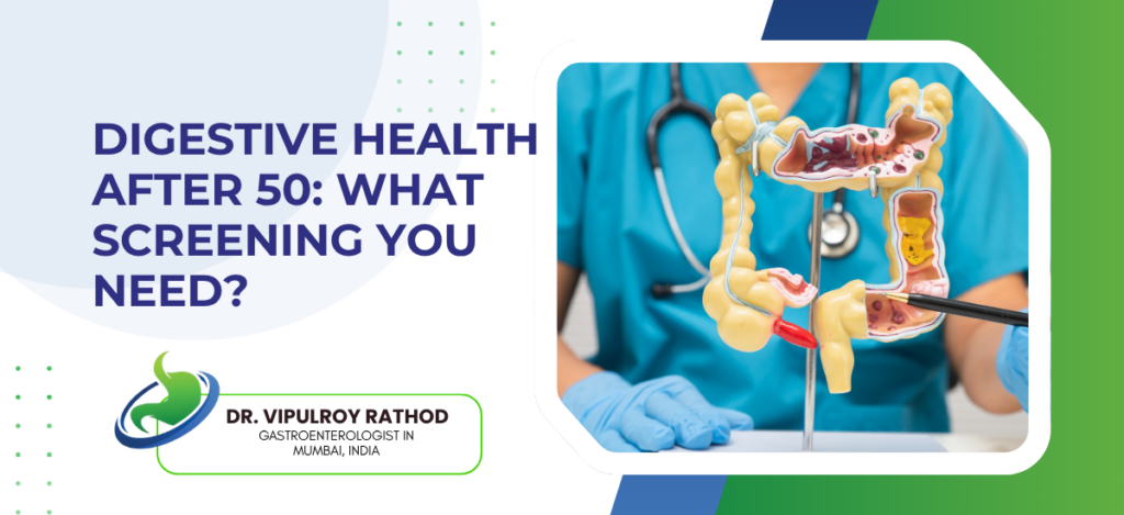
Digestive Health After 50: What Screening You Need?

Turning 50 is the age at which several digestive system cancers and chronic conditions start becoming meaningfully more common, and the screening landscape shifts from optional to standard practice. Colorectal cancer is the headline reason, but it isn’t the only one. Liver disease, gallbladder pathology, and upper GI cancers all rise in incidence from this decade onwards. Most patients only get tested once symptoms appear, which is often too late for the kind of early-stage disease that screening exists to catch. According to Dr. Vipulroy Rathod, Gastroenterologist in Mumbai, “The whole point of screening is to catch something before it announces itself. Colorectal cancer at stage one has a survival rate above ninety percent. At stage four it falls to under fifteen. The difference between those two numbers is usually a colonoscopy that happened, or didn’t, in someone’s fifties. Patients underestimate how much that one test changes the trajectory.” Which screening tests actually matter after 50? Not every test marketed as a wellness package is worth doing. The screening that genuinely changes outcomes falls into a handful of clear categories. Colonoscopy. The single most important GI screening test after 50. One colonoscopy with no polyps means the next one can usually wait ten years. Polyps found and removed reset that schedule, but they also remove the precursor lesions that would have become cancer. This is the test most likely to save a life in this age group. Upper GI endoscopy is selectively useful, not routine for everyone. Patients with longstanding reflux, Indian patients with risk factors for stomach cancer, or anyone with persistent dyspepsia, anaemia, or weight loss should have one. Routine universal upper GI screening isn’t recommended even after 50 unless risk factors are present. FibroScan and liver assessment. Metabolic liver disease affects nearly a third of urban Indian adults over 50, and most patients have no idea. A FibroScan with basic blood tests stages the disease properly and identifies which patients need active management versus reassurance. Stool-based tests as adjuncts, not replacements. FIT (faecal immunochemical test) annually is reasonable for patients who decline colonoscopy outright. Stool DNA tests are available too but currently underused in India. Neither is as sensitive as colonoscopy, both miss meaningful disease. What patients commonly skip is the assessment that builds on the screening results. Endoscopy in Mumbai services that combine colonoscopy and upper GI endoscopy with FibroScan and metabolic workup in the same appointment slot make screening practical for working adults who can’t take time off repeatedly for separate tests. What are the Risk factors that change the screening schedule? Standard screening intervals are designed for average-risk patients. Specific risk factors shift the schedule earlier, more frequently, or to different tests entirely. The first situation is family history. A first-degree relative with colorectal cancer means screening should start ten years earlier than their age at diagnosis, or at 40, whichever comes first. Two or more affected relatives, or any with diagnosis under 50, raises the bar to genetic counselling and consideration of inherited syndromes like Lynch syndrome. The second is personal history of polyps, inflammatory bowel disease, or previous GI cancer. These patients are on surveillance schedules rather than standard screening intervals, often with colonoscopy every one to three years depending on the previous findings. The third is metabolic risk. Diabetes, obesity, fatty liver, and metabolic syndrome significantly raise the risk of multiple GI cancers and chronic liver disease. These patients benefit from earlier and more comprehensive screening, including FibroScan, lipid profile, and HbA1c alongside the standard tests. Finally, there are the symptom-driven indications that aren’t really screening at all, they’re diagnostic workup. Any patient with unexplained weight loss, anaemia, persistent change in bowel habit, rectal bleeding, or significant dyspepsia after 50 needs full GI evaluation regardless of when their last screening was. Read more on colonoscopy and what it detects for how the most important single test in this group is actually performed and interpreted. Why choose Dr. Vipulroy Rathod for digestive health screening? Dr. Vipulroy Rathod has been performing GI screening, surveillance, and complex endoscopy at Fortis Hospital Mulund for over three decades, with the volume of colonoscopies and upper GI endoscopies that builds the pattern recognition needed to spot early disease. The screening agenda for any individual patient gets tailored to family history, metabolic risk, and previous findings rather than applied as a generic protocol. The bigger issue most patients face isn’t access to screening, it’s getting the right tests rather than the wrong ones. Some arrive having spent significantly on full-body checkup packages that didn’t include a colonoscopy. Others arrive after years of stool tests when their family history warranted a scope from the start. Matching the test to the patient is what makes screening actually work. Book your consultation today with one of India’s most experienced specialists for digestive health screening after 50. Book Appointment Call now Frequently Asked Questions At what age should colonoscopy screening start? Standard average-risk screening starts at 45 to 50 depending on guideline. Patients with family history or other risk factors should start earlier, sometimes as young as 40. How often should screening be repeated? A normal colonoscopy with no polyps usually means the next one is in ten years. Polyps found, IBD, or family history shorten the interval considerably. Are stool tests good enough on their own? Better than no screening, not as good as colonoscopy. FIT misses around twenty to thirty percent of significant polyps and a meaningful proportion of cancers. Reasonable for patients who refuse a scope, not the first choice. What other screenings besides colonoscopy matter after 50? Upper GI endoscopy when symptoms or risk factors are present. FibroScan for metabolic liver disease in patients with obesity, diabetes, or dyslipidaemia. Routine ultrasound for the gallbladder in selected patients with risk factors. Reference links- Colorectal Cancer Screening Guidelines, American College of Gastroenterology — https://gi.org/guideline/colorectal-cancer-screening/ Cancer Screening Recommendations, World Health Organization — https://www.who.int/health-topics/cancer

Digestive Health After 50: What Screening You Need? Read More »

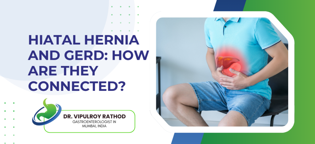
Hiatal Hernia and GERD: How Are They Connected?

A hiatal hernia is when part of the stomach pushes up through the diaphragm into the chest, and GERD is the acid reflux that often comes with it, though not always. The two conditions are linked but they aren’t the same thing. Plenty of patients have hiatal hernias without any reflux symptoms. Plenty have severe GERD without any hernia. When they do coexist, which is common, the hernia makes the reflux significantly harder to control because the anti-reflux barrier itself has been mechanically disrupted. According to Dr. Vipulroy Rathod, Gastroenterologist in Mumbai, “The mistake patients make is thinking hiatal hernia and GERD are the same diagnosis. They’re not. Some hernias cause symptoms, most don’t. What matters is whether the lower oesophageal sphincter is still working. A small sliding hernia in a patient with no reflux needs nothing. A large hernia with severe acid breakthrough needs proper assessment and sometimes surgery.” How a hiatal hernia disrupts the anti-reflux barrier? The lower oesophagus has a built-in anti-reflux mechanism, the lower oesophageal sphincter (LES) muscle, plus the diaphragm pinching the area just above where the stomach becomes oesophagus. Both elements need to be in the right anatomical position to work. Sliding hiatal hernia (Type I). The most common type, accounting for around ninety percent of hernias. The gastro-oesophageal junction slides upwards into the chest, separating it from the diaphragmatic pinch, and the sphincter mechanism is weakened. Reflux risk rises with hernia size. Paraoesophageal hernias (Types II, III, IV) are anatomically more complex and less commonly associated with classical reflux symptoms. The gastro-oesophageal junction often stays below the diaphragm, but other parts of the stomach herniate alongside, and these can present with chest pain, anaemia, or obstructive symptoms rather than acid reflux. Mechanical disruption of the crural diaphragm. A normal diaphragm wraps tightly around the junction and acts as a secondary anti-reflux barrier. A hernia opens this up, and the support system meant to back up the sphincter is no longer there to do its job during coughing, lifting, or lying flat. Acid pocket effect. In hiatal hernia patients, gastric acid sometimes pools above the diaphragm in a small pocket that sits closer to the oesophagus than normal, leading to higher acid exposure even when overall reflux frequency isn’t dramatically increased. The clinical impact depends on the size of the hernia and how badly the sphincter is functioning. GERD treatment approaches in patients with hiatal hernia often need to address both elements, the acid suppression and the mechanical defect, because PPIs alone may not control symptoms once the anti-reflux barrier has been physically broken. When the connection matters clinically? Not every patient with both conditions needs aggressive treatment, but knowing the connection changes how the workup proceeds. The first situation where it matters is in patients who haven’t responded adequately to PPI therapy. Around thirty to forty percent of GERD patients have persistent symptoms despite proper PPI dosing, and a significant proportion of these have a substantial hiatal hernia that’s making medical treatment inadequate. The second is in patients being considered for anti-reflux surgery. Hiatal hernia repair is now an integral part of fundoplication, and surgeons want detailed assessment of hernia size, type, and anatomy before any operation. Without proper hernia repair, fundoplication often fails within a few years. The third situation is in older patients with paraoesophageal hernias and atypical symptoms. Chest pain mimicking cardiac disease. Iron deficiency anaemia from a Cameron’s ulcer at the diaphragmatic constriction. Postprandial fullness and early satiety. These patients often go unrecognised because their symptoms don’t fit the classical reflux pattern. Diagnosis usually relies on endoscopy, sometimes with manometry and pH studies when surgery is being considered, and imaging in selected cases. Read more on what GERD is and when treatment is needed for how reflux disease is assessed and treated in patients with or without an associated hernia. Why choose Dr. Vipulroy Rathod for hiatal hernia and GERD assessment? Dr. Vipulroy Rathod has been managing GERD, hiatal hernia, and the overlap between them at Fortis Hospital Mulund for over three decades. The assessment of these patients combines endoscopy, pH studies where relevant, manometry, and the surgical coordination needed for patients whose reflux can’t be controlled with medical treatment alone. The hardest cases aren’t the obvious ones. The hardest are patients with subtle anatomy, atypical symptoms, or partial PPI response, where the question of whether the hiatal hernia is contributing significantly determines whether surgery is the right step. Getting that decision right requires looking at the whole picture rather than treating either condition in isolation. Book your consultation today with one of India’s most experienced specialists for hiatal hernia and GERD assessment. Book Appointment Call now Frequently Asked Questions Does every hiatal hernia cause reflux? No. Most small sliding hernias cause no symptoms. The connection becomes clinically relevant when the hernia is large enough to disrupt the anti-reflux barrier significantly. Can a hiatal hernia be fixed without surgery? Not the hernia itself, no. Surgery is the only definitive repair. Symptoms from a hernia, including reflux, can often be controlled medically without ever needing operative intervention. When does hiatal hernia need surgery? Mainly in two situations. Severe GERD not controlled by PPIs. Or large paraoesophageal hernias causing chest pain, anaemia, or obstructive symptoms, regardless of reflux. Can fundoplication fail without proper hernia repair? Yes, and this is one of the commonest reasons for fundoplication failure. Modern anti-reflux surgery always includes hiatal hernia repair as part of the procedure for this reason. Reference links- GERD Clinical Guidelines, American College of Gastroenterology — https://gi.org/guideline/diagnosis-and-management-of-gastroesophageal-reflux-disease/ Hiatal Hernia Management, Society of American Gastrointestinal and Endoscopic Surgeons — https://www.sages.org/publications/guidelines/guidelines-for-the-management-of-hiatal-hernia/

Hiatal Hernia and GERD: How Are They Connected? Read More »

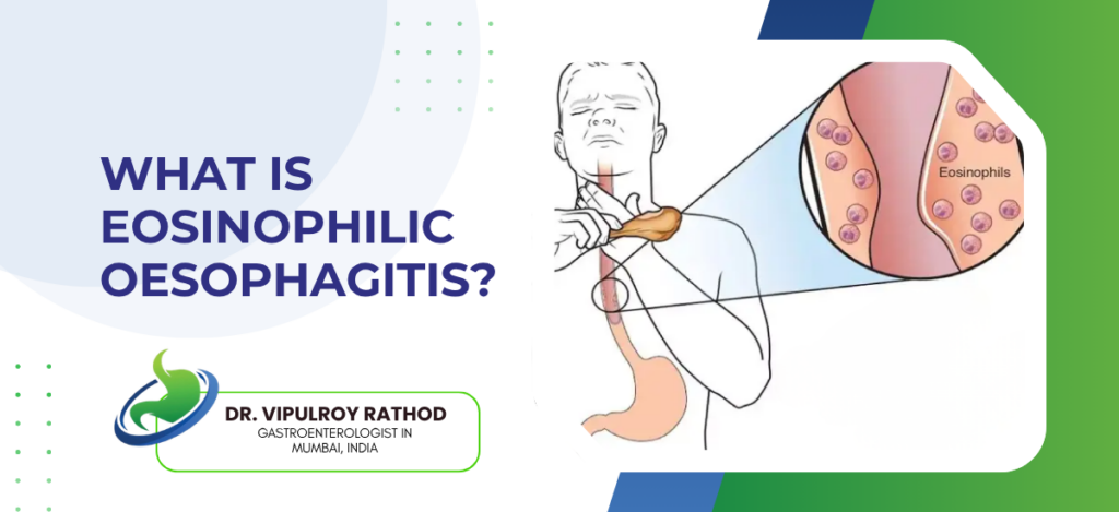
What Is Eosinophilic Oesophagitis?

Eosinophilic oesophagitis (EoE) is a chronic, allergy-driven inflammation of the oesophagus where eosinophils, a type of white blood cell, build up in the lining and cause swelling, scarring, and difficulty swallowing. It is increasingly recognised as a major cause of dysphagia and food impaction in younger adults. The condition often gets misdiagnosed as reflux for years before someone takes a biopsy and counts the eosinophils, which is the only way to actually make the diagnosis. EoE isn’t life-threatening, but untreated it progressively narrows the oesophagus and reduces quality of life significantly. According to Dr. Vipulroy Rathod, Gastroenterologist in Mumbai, “Patients with EoE often come to us after years of being treated for reflux that never quite settled. They’ve tried PPIs, lifestyle changes, sometimes even surgery for hiatus hernia. The clue is usually food sticking, particularly meat and bread, in young or middle-aged patients. Once we look properly with biopsy, the diagnosis becomes obvious.” How EoE develops and how it’s diagnosed? EoE is allergy-driven but the allergy lives in the food pipe rather than anywhere else, which is what makes it so easy to miss. Food triggers, mostly. Cow’s milk and wheat lead the list. Eggs, soy, and nuts after that. Some patients also have aeroallergen triggers, pollen and dust mites, especially if they have asthma or eczema in the background. The symptom that should make any doctor think of EoE is solid food sticking. Bread, dry meat, rice. The patient stops, drinks water, the food eventually clears. Many patients have developed elaborate coping strategies over years, obsessive chewing, drinking water with every bite, avoiding certain foods entirely, without ever realising they’re managing an undiagnosed disease. Endoscopy with biopsy is non-negotiable. Looking at the oesophagus alone tells you something. Rings, furrows, white plaques, narrowing. But the diagnosis only becomes official when biopsies from multiple levels show more than fifteen eosinophils per high-power field on the histology report. Allergy blood tests and skin prick testing? Less useful than they sound. They don’t reliably predict which foods are driving any individual patient’s disease, and elimination diets work better when designed by trial rather than by lab result. Getting EoE diagnosed properly changes the management completely. Endoscopy in Mumbai services should be the first stop for anyone with persistent dysphagia or food impaction, because the multi-level biopsies needed to confirm or rule out EoE can’t be done without the right scope and a willingness to biopsy even a normal-looking oesophagus. How EoE is treated? Three angles to treatment. Calm the inflammation, find and remove the trigger food, open up any narrowing that’s already developed. Topical steroids. Swallowed budesonide or fluticasone, formulated to coat the oesophagus rather than get absorbed into the bloodstream. Works in most patients. Response confirmed at eight to twelve weeks with repeat biopsy. High-dose proton pump inhibitors actually treat a meaningful subset of EoE patients on their own. The mechanism isn’t only acid suppression, PPIs have a direct anti-inflammatory effect on eosinophils that wasn’t recognised for years. Around thirty to forty percent of patients respond to PPI monotherapy. Elimination diets. Six-food elimination is the classical approach, removing the six commonest trigger food groups, then reintroducing them one by one with repeat endoscopy to identify the actual culprit. Four-food and two-food versions exist for patients who can’t tolerate the full elimination. Endoscopic dilatation for established strictures. EoE patients sometimes develop tight rings or fixed narrowings after years of inflammation, and dilatation reopens them mechanically. Always combined with continued medical treatment to prevent the inflammation from coming back. So treatment is rarely one thing. Read more on reflux vs other GI Conditions for how EoE fits into the differential diagnosis of long-standing oesophageal symptoms. Why choose Dr. Vipulroy Rathod for EoE diagnosis and management? Dr. Vipulroy Rathod has been diagnosing and managing eosinophilic oesophagitis at Fortis Hospital Mulund for over three decades, and the difference experience makes here is mostly about recognition. EoE looks subtle on endoscopy, the rings and furrows are easy to dismiss as normal variation, and without a low threshold for biopsying patients with dysphagia or food impaction, the diagnosis gets missed. Patients arrive having had multiple scopes elsewhere where the diagnosis wasn’t even considered, sometimes after years of PPI treatment for reflux that wasn’t actually reflux, occasionally after hiatus hernia surgery that didn’t fix the problem because the problem was never reflux to begin with. Book your consultation today with one of India’s most experienced specialists for swallowing problems and EoE assessment. Book Appointment Call now Frequently Asked Questions Is EoE the same as acid reflux? No. Different cause, different treatment, but they can coexist in the same patient. How is EoE confirmed? Endoscopy plus biopsies from multiple oesophageal levels. More than fifteen eosinophils per high-power field is the threshold. Can EoE be cured? Controlled, not cured. Stopping treatment usually means the inflammation returns within months. Is EoE dangerous? Not in the short term. Long-term, untreated EoE narrows the oesophagus and causes recurrent food impactions, which is a real quality-of-life problem and occasionally an emergency. Reference links- Eosinophilic Oesophagitis Guidelines, American College of Gastroenterology — https://gi.org/guideline/eosinophilic-esophagitis/ AGREE Conference Updates on EoE, World Gastroenterology Organisation — https://www.worldgastroenterology.org/guidelines

What Is Eosinophilic Oesophagitis? Read More »

Pancreatic Cancer Pain: How Is It Managed?

Pancreatic cancer pain is among the most severe pain encountered in oncology, caused by tumour invasion of the celiac plexus, ductal obstruction, and inflammation in surrounding tissues. Management uses a layered approach, starting with non-opioid analgesia, moving through opioid medication, and adding interventional procedures like EUS-guided celiac neurolysis when standard treatment isn’t enough. The goal isn’t only pain reduction. It is functional quality of life, the ability to eat, sleep, and spend time meaningfully. Many patients have their pain undertreated for months before someone takes it seriously enough to escalate appropriately. According to Dr. Vipulroy Rathod, Gastroenterologist in Mumbai, “Pancreatic cancer pain is different from most other pains, and treating it like it’s the same as back pain or arthritis is where things go wrong. It needs early opioid use, often combined with adjuvants, and procedural pain blocks much earlier than they tend to be offered. Patients shouldn’t have to wait until the pain is unbearable before someone tries something different.” What are the layered approach to pain management? Pancreatic cancer pain isn’t controlled by any single medication or procedure. It needs several treatments working alongside each other, escalating as the disease progresses. Non-opioid analgesia as the baseline. Paracetamol and NSAIDs in the early stages, though NSAIDs are limited by kidney function and bleeding risk in many cancer patients. Useful when the pain is still moderate. Opioid medication is started earlier in pancreatic cancer than in most other conditions. Oral morphine, oxycodone, or fentanyl patches, with breakthrough doses available for flare-ups. The fear of starting opioids early is misplaced in this disease, the issue is usually undertreatment, not addiction. Adjuvant medications. Gabapentin or pregabalin for the neuropathic component, tricyclic antidepressants in some cases, and corticosteroids for short-term pain flares. These often allow lower opioid doses than would otherwise be needed. EUS-guided celiac plexus neurolysis for pain that doesn’t respond to medical management. Alcohol injection around the celiac plexus permanently interrupts pain signals from the upper abdomen, with relief lasting weeks to months and often the rest of the patient’s illness. Choosing the right combination depends on where the patient is in their disease and how much function they still have. Pancreatic cancer treatment planning at every stage should include pain management as part of the discussion, not an afterthought once everything else has been settled. What changes through the disease course? Pain management isn’t static. The drugs that worked early often stop working, and what’s needed evolves with the disease. Early disease, mild-moderate pain. Paracetamol, NSAIDs where safe, low-dose opioids for breakthrough. Many patients are managed on relatively modest medication during the first few months after diagnosis if chemotherapy is shrinking the tumour or controlling growth. As the disease progresses, opioid doses usually need to rise. Pain that was manageable on twenty milligrams of morphine a day might need eighty or more six months later. Rotation between different opioids is often used when tolerance develops to one specific drug. The point where celiac neurolysis should be considered. Patients on rising opioid doses with breakthrough pain, side effects from medication, and a quality of life that’s eroding because of the pain itself, not just the cancer. This is usually weeks or months earlier than it actually gets offered in most centres. End-of-life pain management. Subcutaneous opioid infusions, syringe drivers for continuous dosing, and palliative care integration. By this stage the focus is comfort and symptom control rather than function. The aim throughout is to keep pain controlled without trading too much of the patient’s alertness or quality of life for that control. Read more on pancreatic cancer detection to understand why so many cases are picked up at stages where pain management becomes a major part of the conversation from the start. Why choose Dr. Vipulroy Rathod for pancreatic cancer pain management? Dr. Vipulroy Rathod has been managing pancreatic cancer pain at Fortis Hospital Mulund for over three decades, with particular focus on EUS-guided celiac plexus neurolysis and the procedural side of pain management that most centres don’t offer routinely. Many patients arrive on rising opioid doses with poor pain control, having never been offered celiac neurolysis as an option earlier in their illness. Pain in this disease deserves the same attention as the cancer itself. Patients spending their last months in unrelieved pain because no one referred them for a celiac block is, frankly, a system failure rather than a clinical limit. The block isn’t experimental or last-resort, it’s a procedure that’s been available for decades and works. Book your consultation today with one of India’s most experienced specialists for pancreatic disease pain management and procedural intervention. Book Appointment Call now Frequently Asked Questions Why is pancreatic cancer pain so severe? The pancreas sits close to the celiac plexus, a major bundle of nerves carrying visceral pain signals. Tumour invasion of this plexus directly produces some of the most intense pain encountered in cancer, often poorly controlled by standard analgesics without procedural intervention. When should celiac plexus neurolysis be considered? Earlier than it usually is. Rising opioid doses, breakthrough pain not controlled by medication, side effects affecting quality of life, all of these are signs the procedure should be on the table. Waiting until pain is unbearable is the common mistake. Are opioids addictive in this setting? Addiction is rarely a meaningful concern in pancreatic cancer pain management. Physical dependence develops, but that’s different from addiction, and the priority is pain control rather than minimising opioid exposure. Undertreatment harms more patients than overuse does. Does pain management affect cancer treatment? No, the two run in parallel. Good pain control actually helps patients tolerate chemotherapy better and stay functional longer, which can improve overall survival. Pain management isn’t a separate track from cancer treatment, it’s part of the same plan. Reference links- EUS-guided celiac plexus neurolysis — https://ascopubs.org/doi/10.1200/JCO.2010.32.2750 (JCO trial on early EUS-CPN in pancreatic cancer) WHO ladder & cancer pain management — https://www.annalsofoncology.org/article/S0923-7534(19)31698-9/fulltext (ESMO Clinical Practice Guidelines)

Pancreatic Cancer Pain: How Is It Managed? Read More »

What Is EUS Guided Celiac Block for Pain?

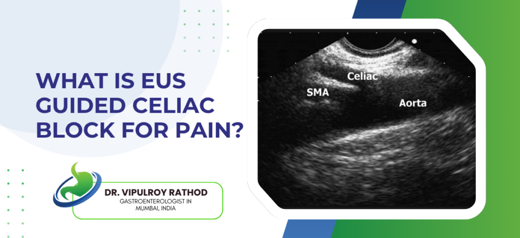
EUS-guided celiac plexus block is an endoscopic procedure that delivers a steroid and local anaesthetic injection, or sometimes a neurolytic agent, directly into the celiac plexus, the bundle of nerves carrying pain signals from the upper abdominal organs. It is used to control severe abdominal pain in patients with chronic pancreatitis and unresectable pancreatic cancer. The procedure replaces the need for escalating opioid doses in many patients, particularly those whose pain has stopped responding to standard medication. Done through endoscopic ultrasound rather than fluoroscopy or CT, the access is more direct and the targeting more precise than older percutaneous techniques. According to Dr. Vipulroy Rathod, Gastroenterologist in Mumbai, “Pancreatic pain is a different category from other abdominal pain. It comes from deep visceral nerves, doesn’t respond well to standard painkillers, and gets worse as the disease progresses. Celiac block targets the wiring directly. It doesn’t fix the cause, but it interrupts the signal, and for the right patient that’s life-changing.” Who benefits from EUS-guided celiac block? The two main indications are chronic pancreatitis and unresectable pancreatic cancer, but the type of injection used differs between them. Chronic pancreatitis with severe pain. Patients whose pain has stopped responding to enzyme replacement, analgesia, and lifestyle changes. The injection here is usually a steroid plus local anaesthetic combination, called a celiac block rather than neurolysis, and the effect lasts weeks to months. Some patients need repeat injections every few months. Unresectable pancreatic cancer pain is where neurolysis becomes appropriate. Absolute alcohol is injected to permanently damage the nerves, and pain control often lasts the rest of the patient’s life. The trade-off is that neurolysis is permanent and can’t be reversed if complications develop. Failure of medical pain management. Patients on rising opioid doses who are getting side effects without proper pain control. Celiac block lets opioid doses come down, often substantially, while pain stays better managed than before. Selected cases of locally advanced disease awaiting chemotherapy response, where pain control is needed urgently while definitive treatment plays out over months. The decision between block and neurolysis depends on the underlying disease and prognosis. Pancreatic cancer treatment planning in advanced disease almost always includes a conversation about pain management options, and celiac block fits early into that conversation rather than being a last resort after everything else has failed. How the procedure works and what to expect? EUS-guided celiac block is done under sedation, usually as a day-care procedure, taking around twenty to thirty minutes from start to finish. A linear-array echoendoscope is passed through the mouth into the stomach, positioned to image the celiac axis and the bundle of nerves surrounding it. Under real-time ultrasound guidance, a fine needle is advanced through the stomach wall to the area just around the celiac plexus. The injection is given, usually divided between two sites on either side of the celiac axis for better coverage. Then the needle is withdrawn. If you’re the patient, the experience is straightforward. You arrive in the morning, sedation goes in, you wake up in recovery an hour later, you go home by afternoon. Pain relief usually starts within twenty-four to forty-eight hours. Block (steroid plus anaesthetic) effects. Pain relief in around sixty to seventy percent of chronic pancreatitis patients, with effects lasting weeks to several months. Repeat injections work but with diminishing returns over multiple cycles. Neurolysis (alcohol injection) effects. Substantial pain relief in seventy to eighty percent of pancreatic cancer patients, with effects often lasting the duration of the illness. Some patients have no effect, and the lack of response is itself information about the source of the pain. Complications are uncommon but possible. Transient diarrhoea, hypotension after the procedure (from sympathetic blockade), occasional retroperitoneal bleed. Permanent paraplegia from spinal cord injury has been reported but is rare in EUS-guided cases compared to older posterior approaches. Repeat blocks are technically straightforward in chronic pancreatitis patients. Neurolysis, once done, is permanent and isn’t repeated. So the choice of agent depends on whether you’re treating a long-term condition or a terminal one. Read more on pancreatic cancer detection for how pain management decisions fit into the broader pathway of unresectable pancreatic disease. Why choose Dr. Vipulroy Rathod for EUS-guided celiac block? Dr. Vipulroy Rathod has been performing EUS-guided celiac block and neurolysis at Fortis Hospital Mulund for over three decades, covering both chronic pancreatitis cases and unresectable pancreatic cancer pain management. Many patients arrive on high doses of opioid medication, struggling with side effects and inadequate pain control, having never been offered celiac block as an option earlier in their disease course. The procedure isn’t a last resort. It works best when offered before pain becomes intractable, before opioid tolerance is fully established, before the patient’s quality of life has eroded too far. Timing matters as much as technique. Book your consultation today with one of India’s most experienced specialists for EUS-guided pain management and pancreatic disease care. Book Appointment Call now Frequently Asked Questions Is EUS-guided celiac block painful? The procedure itself isn’t, because it’s done under sedation. Some patients have transient abdominal discomfort for a day or two afterwards, which settles on its own. Severe pain after the procedure is uncommon and would need assessment. How long does the pain relief last? For chronic pancreatitis blocks, several weeks to several months, often with diminishing duration on repeat injections. For pancreatic cancer neurolysis, often the rest of the patient’s illness, though some patients need a second procedure if relief fades. Can the procedure be repeated? Steroid-based blocks for chronic pancreatitis can be repeated several times. Neurolysis with alcohol is generally a one-time procedure, though there are rare cases where it’s repeated when partial response occurred initially. What are the side effects? Diarrhoea and a drop in blood pressure are the most common, both usually mild and self-limiting. More serious complications like retroperitoneal bleeding or, very rarely, spinal cord injury, are uncommon in experienced hands using EUS guidance. Reference links- EUS-Guided Therapeutic Procedures Guidelines, American Society for Gastrointestinal Endoscopy —

What Is EUS Guided Celiac Block for Pain? Read More »

When Is a Pancreatic Stent Needed?

A pancreatic stent is needed when the pancreatic duct is blocked, narrowed, or leaking, and that obstruction is causing pain, recurrent pancreatitis, or interfering with drainage. The decision isn’t automatic. Some duct narrowings can be watched, others need urgent intervention, and the difference depends on symptoms, imaging, and whether the cause is something correctable on its own. Most stents go in during ERCP, sometimes with EUS guidance for difficult anatomy. The harder question for most patients isn’t whether stenting works, it’s whether they actually need one right now. According to Dr. Vipulroy Rathod, Gastroenterologist in Mumbai, “Stenting is overused in some centres and underused in others. A patient with mild chronic pancreatitis and minor duct changes doesn’t always need a stent. A patient with a tight stricture and recurrent pain definitely does. Getting that judgment right is what separates good pancreatic ERCP practice from procedural overuse.” Which are the Conditions that typically need stenting? The indications fall into a handful of clear categories, and most cases that get stented fall into one of these patterns. Dominant strictures in chronic pancreatitis. A localised narrowing of the main duct, often with upstream dilatation visible on MRCP, in a patient with ongoing pain. Stenting the stricture relieves pressure and pain in around sixty to seventy percent of cases. Pancreatic duct stones, particularly when they’re impacted and causing recurrent pancreatitis. Removal comes first, sometimes with shockwave lithotripsy if the stones are large or hard, then the stent goes in to keep the duct open while the inflammation calms down. Pancreatic duct leak or disruption. Trauma, surgery, or severe pancreatitis can tear the duct. Pancreatic juice then leaks into surrounding tissue, sometimes forming pseudocysts or causing ascites. A transpapillary stent across the leak point usually allows it to heal. Some pancreatic cancers compressing the duct. Not all of them, only the small subset where ductal pressure is causing pain or recurrent pancreatitis and surgery isn’t an option, often in patients on chemotherapy who need symptom control. Choosing which patients benefit isn’t always obvious from imaging alone. Pancreatic stone extraction work and stenting decisions usually happen together in the same procedure, with EUS sometimes added beforehand to assess the duct anatomy, exclude masses, and plan the approach properly. When stenting is the wrong answer? Not every blocked duct should be stented, and the reverse question matters as much as knowing when to do it. Asymptomatic minor duct changes. Mild narrowing on MRCP without pain, without recurrent pancreatitis, without functional impairment. Stenting these patients exposes them to procedure risk without clear benefit. Acute pancreatitis episodes themselves aren’t an indication for stenting in most cases. The duct usually clears on its own once inflammation settles. Premature stenting can make things worse rather than better. Diffuse strictures throughout the entire pancreatic duct. When the whole duct is narrowed rather than one focal segment, stenting one area doesn’t help much, and these patients often need surgical drainage rather than endoscopic stenting. Patients who can’t commit to the follow-up schedule. A stent left in for years causes more problems than it solved, and patients who can’t return for exchange every three to six months are often better managed with non-stenting strategies like enzyme replacement and pain management. You don’t want a stent placed because something can be stented. You want one placed because the alternative was worse. Read more on pancreatic stenting for how the procedure itself works once that decision has been made. Why choose Dr. Vipulroy Rathod for pancreatic stenting decisions? Dr. Vipulroy Rathod has been managing pancreatic ERCP and stenting decisions at Fortis Hospital Mulund for over three decades, with particular focus on chronic pancreatitis where the question of who benefits from stenting is harder than it looks. Many patients arrive having been told they need urgent stenting based on imaging alone, when a closer look at their symptoms and pain pattern suggests they don’t. Others arrive having been managed non-procedurally for years when a single well-timed stent would have changed their pain trajectory significantly. Knowing when not to stent is the harder skill. The procedure itself isn’t trivial, the complication rate isn’t zero, and the follow-up commitment is real. Patients who understand all of that before the first procedure tend to do better than those who weren’t told upfront. Book your consultation today with one of India’s most experienced specialists for pancreatic disease assessment and stenting decisions. Book Appointment Call now Frequently Asked Questions Does every chronic pancreatitis patient need a stent? No. Many are managed perfectly well with enzyme replacement, pain medication, and lifestyle changes. Stenting is reserved for patients with dominant strictures, recurrent pancreatitis, or pain not controlled by conservative measures. How urgently does a pancreatic stent need to be placed? It depends. Pancreatic duct leak after trauma needs urgent stenting within days. A dominant stricture causing chronic pain can be planned electively. Acute pancreatitis usually doesn’t need urgent stenting at all. Can a pancreatic stent be avoided with medication alone? For some patients yes, particularly those with milder duct changes and pain controlled by analgesia and enzyme replacement. For tight strictures causing recurrent pancreatitis, medication alone usually isn’t enough. Is stenting permanent? No. Plastic stents are exchanged every three to six months, and most patients eventually have them removed entirely once the underlying problem stabilises. Long-term stenting beyond a year or two is uncommon and usually means the underlying condition needs reassessment. Reference links- ERCP and Pancreatic Stenting Guidelines, American Society for Gastrointestinal Endoscopy — https://www.asge.org/home/practice-support/guidelines Chronic Pancreatitis Management Standards, World Gastroenterology Organisation — https://www.worldgastroenterology.org/guidelines  

When Is a Pancreatic Stent Needed? Read More »

Scroll to Top
Call Now Button