Dr. Vipulroy Rathod

Author name: Dr. Rathod Medical Foundation

Banner for an article: 'Ulcerative Colitis While Pregnant' with a detailed gut diagram and a magnified inflamed tissue view, plus the doctor’s logo and name.

Can You Have a Healthy Pregnancy With Ulcerative Colitis?

Ulcerative Colitis While Pregnant: What You Need to Know A pregnancy diagnosis, planned or unplanned, changes the questions a person with ulcerative colitis is suddenly asking themselves. Will the disease flare again once hormones shift? Is it safe to keep taking the same medication? Can the pregnancy even be carried to term without complications? For the large majority of patients, the answer is reassuring: a healthy pregnancy is very much achievable, provided the disease is planned around rather than left to chance. Dr. Vipulroy Rathod, a globally acclaimed gastroenterologist in Mumbai, India, has spent more than three decades treating inflammatory bowel disease, including ulcerative colitis, and has walked many patients through this exact life stage. In his experience, the single biggest predictor of how a pregnancy unfolds is not the diagnosis itself but the state of the disease at the moment of conception. Patients who become pregnant while their disease is quiet tend to stay that way; patients who conceive mid-flare are more likely to see that flare persist or worsen. At his practice in Mumbai, Dr. Rathod pairs advanced endoscopic evaluation with close coordination alongside each patient’s obstetrician, so that ulcerative colitis management and pregnancy care are never working against each other. His approach to inflammatory bowel disease treatment in Mumbai is built around exactly this kind of long-term, life-stage planning rather than treating flares in isolation. Does Ulcerative Colitis Affect Fertility and Pregnancy? Ulcerative colitis on its own does not meaningfully lower a person’s chances of conceiving, particularly when the disease is inactive. Fertility during remission tends to run close to that of the general population. There is one notable exception: surgery that creates an internal pouch from the small intestine, sometimes needed in more severe or long-standing disease, can reduce fertility because of scarring around the pelvis. Anyone considering this type of surgery should raise fertility as part of that conversation, ideally before the procedure rather than after. Pregnancy development and progression is closely linked to the severity and extent of the disease after a positive conception takes place. A flare of the condition either at the time of conception or later during pregnancy A lot increases chances of an early delivery, birth of a baby with lower than normal weight, and even miscarriage according to the seriousness of the situation. However, interestingly that women with active ulcerative colitis who go on to conceive and carry baby to term have an increased risk of complications during pregnancy, but this does not appear to be a consequence of Really they are pregnant but rather results of continued inflammation. Planning a Pregnancy With Ulcerative Colitis Time conception around remission. A period of stable, medication-controlled remission lasting three to six months before trying to conceive gives the best odds of a settled pregnancy. Have your medications reviewed early. A few drugs, methotrexate being the clearest example, need to be stopped well ahead of conception, while most maintenance treatments can continue without interruption. Check nutritional status. Iron, folate, vitamin D, and vitamin B12 are worth testing and correcting beforehand, since bowel inflammation can interfere with how well these nutrients are absorbed. Bring your obstetrician and gastroenterologist together early. When both specialists are aligned from the outset, patients are far less likely to receive conflicting advice partway through pregnancy. Resist the urge to stop treatment out of caution. Quietly discontinuing medication for fear of harming the baby is, ironically, one of the more common triggers for a flare during pregnancy. Which Ulcerative Colitis Medications Are Considered Safe During Pregnancy? Few questions cause more anxiety among pregnant patients than this one, and unfortunately, a lot of outdated or overly cautious information still circulates. The broader clinical picture is worth holding onto: uncontrolled ulcerative colitis poses a greater risk to a pregnancy than most of the standard treatments used to control it. Aminosalicylate medications: These are widely regarded as safe to continue throughout pregnancy and while breastfeeding. Corticosteroids: Used when a flare needs to be brought under control quickly, generally at the lowest dose that works and for as short a period as possible. Biologic medications, including tumour necrosis factor inhibitors, vedolizumab, and ustekinumab: Data from large patient registries has not shown a meaningful rise in pregnancy complications or early childhood problems, so most specialists now continue these medications through pregnancy for patients who need them to stay in remission. Thiopurines such as azathioprine and mercaptopurine: Typically continued in patients who are already stable on them, since withdrawing the medication can bring the disease back. Methotrexate: Not used in pregnancy under any circumstance, and stopped several months before conception because of a clearly established risk of birth defects. Janus kinase inhibitors and other newer oral medications: Long-term pregnancy data for these is still limited, so specialists generally switch patients to a better-studied option before they try to conceive. One detail patients often miss: the timing of certain biologic doses in the final trimester can influence which vaccines a newborn should avoid in the first months of life. That’s a detail worth confirming directly with your treating specialist rather than piecing together from general reading. Not sure whether your current treatment plan is appropriate for pregnancy? Get in touch with Dr. Rathod’s team at Fortis Hospital, Mulund, before making any changes on your own. Book Appointment Call now What Happens If Ulcerative Colitis Flares During Pregnancy? Flares don’t pause for pregnancy, and when one happens, it needs proper treatment rather than being written off as a normal part of expecting. This can be tricky, since symptoms like more frequent bowel movements or tiredness overlap with ordinary pregnancy changes, which sometimes delays a proper diagnosis. Any new or worsening rectal bleeding, abdominal pain, or fever during pregnancy deserves prompt attention rather than a wait-and-see approach. Investigations are chosen with pregnancy in mind. A limited flexible sigmoidoscopy is generally considered low-risk when clinically necessary, while a full colonoscopy or scans involving radiation are reserved for situations where the information genuinely cannot be obtained another way. A

Can You Have a Healthy Pregnancy With Ulcerative Colitis? Read More »
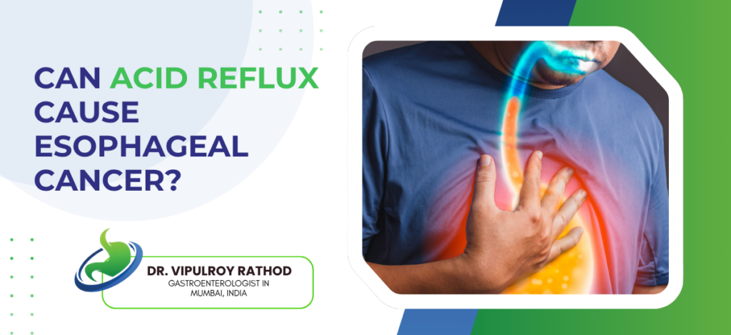
Informational banner asking if acid reflux can cause esophageal cancer, featuring a chest image and doctor Dr. Vipulroy Rathod's logo and title.

Can Acid Reflux Cause Esophageal Cancer?

Acid reflux is among the most common digestive complaints, typically experienced as a burning discomfort after meals. While occasional reflux is harmless, persistent and untreated reflux over several years is recognised as a contributing risk factor for esophageal cancer. Dr. Vipulroy Rathod explains, “Occasional heartburn is rarely a cause for concern. The risk arises from chronic acid reflux that remains unmanaged for years, as the resulting repeated damage to the esophageal lining can gradually lead to serious complications.” Dr. Vipulroy Rathod is a distinguished gastroenterologist in Mumbai, India, with over 30 years of experience and more than 80,000 endoscopic procedures to his credit. A pioneer of diagnostic and interventional endoscopic ultrasound in South Asia, he is widely recognised for detecting esophageal and digestive conditions early, often before symptoms turn serious. His deep expertise in upper GI endoscopy makes him a trusted authority for patients whose reflux needs more than just routine medication. So how does a common complaint like heartburn turn into something serious? Let’s explore the connection step by step. What Is Acid Reflux and How Does It Affect the Esophagus? Acid reflux happens when stomach acid flows backward into the esophagus, the tube that carries food from your mouth to your stomach. A muscular valve called the lower esophageal sphincter normally keeps acid where it belongs. When this valve weakens or relaxes at the wrong time, acid escapes upward and irritates the delicate esophageal lining. That irritation is what causes the familiar burning sensation known as heartburn. An occasional episode is harmless and clears on its own. But when reflux becomes frequent, it is classified as GERD, or gastroesophageal reflux disease. Unlike the stomach, the esophagus has no natural protective coating against acid, so repeated exposure gradually inflames and damages its lining over time. Wondering whether your reflux has crossed from normal to chronic? Let’s dive into what happens when it goes untreated for years. How Can Chronic Acid Reflux Lead to Esophageal Cancer? The link is not immediate, but it is real. When acid repeatedly washes over the esophageal lining for years, the body responds by trying to protect itself. The normal cells lining the esophagus start to change into a different, more acid-resistant type of cell. This adaptation may sound helpful, but it is the first step on a path that can turn dangerous. Over time, these altered cells can become abnormal, a condition doctors call dysplasia. In a small number of people, dysplasia progresses further and develops into esophageal cancer, specifically a type called adenocarcinoma. The key point is that this process unfolds slowly and silently, often over many years, which is exactly why long-standing GERD should never be ignored. The good news is that catching these changes early, before cancer develops, is entirely possible with the right monitoring. But what exactly are these cell changes called, and why do they matter so much? Let’s discuss the condition that sits right in the middle of this journey. What Is Barrett’s Esophagus and Why Does It Matter? Barrett’s esophagus is the name given to that change in the esophageal lining caused by long-term acid exposure. In this condition, the flat cells that normally line the esophagus are replaced by cells similar to those found in the intestine. It develops in a portion of people with chronic GERD and is considered the most significant risk factor for esophageal adenocarcinoma. Here is why it matters. Barrett’s esophagus itself usually causes no extra symptoms, so a person may feel their reflux is under control while the lining quietly continues to change. Most people with Barrett’s never develop cancer, but the condition does raise the risk, which makes regular monitoring essential. Detecting Barrett’s early allows doctors to track it closely and act long before any cancer has a chance to form. This is one of the strongest reasons why persistent reflux deserves a proper look inside with an endoscopy rather than years of guessing from symptoms alone. How do you know if your reflux is heading in this direction? Let’s look at the warning signs worth paying attention to Warning Signs That Acid Reflux May Be Turning Serious Most reflux is manageable, but certain symptoms signal that it is time to see a specialist without delay: Difficulty or pain when swallowing food or liquids A sensation of food getting stuck in the chest or throat Unexplained weight loss with no change in diet Persistent vomiting or vomiting blood Black or tarry stools, which can indicate bleeding A chronic cough, hoarse voice, or sore throat that won’t clear up Heartburn that no longer responds to usual medication Some of these overlap with silent GERD symptoms that don’t feel like typical heartburn at all, which is why they are so easily missed. Noticing any of these signs? Connect with a gastroenterologist before brushing it off as ordinary reflux. Book Appointment Call now Who Is at Higher Risk of Developing Esophageal Cancer? Some people carry a greater risk than others. Knowing where you stand helps you stay ahead of the disease. People with long-standing GERD lasting more than five years Those diagnosed with Barrett’s esophagus Men, who are affected more often than women Adults over the age of 50 Smokers and heavy alcohol users People who are overweight or obese, especially with belly fat Those with a family history of esophageal cancer or Barrett’s People with a diet low in fruits and vegetables How Is the Risk Diagnosed and Monitored? Catching changes early relies on looking directly at the esophagus rather than guessing from symptoms. Here are the main tools doctors use: Upper endoscopy  A thin, flexible tube with a camera is passed into the esophagus to view the lining in real time. It reveals inflammation, Barrett’s changes, and early cancer that symptoms alone cannot show. This is often the moment GERD needs endoscopy rather than just more medicines. Biopsy Small tissue samples are taken during the endoscopy and examined under a microscope. This confirms whether cell changes or dysplasia are present.

Can Acid Reflux Cause Esophageal Cancer? Read More »

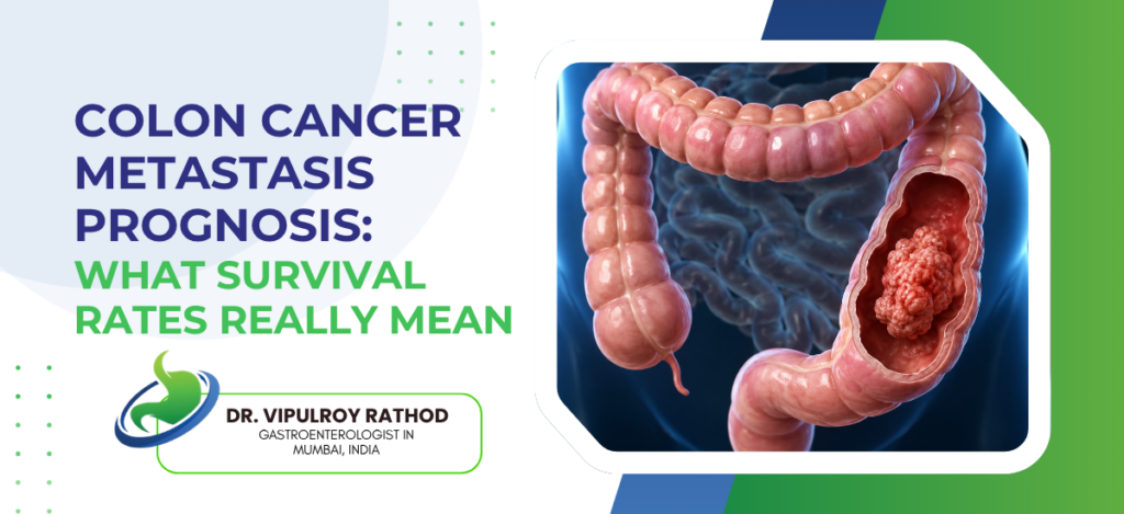
Educational banner on colon cancer prognosis with bold blue/green title and a 3D colon illustration on the right, plus a doctor’s logo area.

Colon Cancer Metastasis Prognosis: What Survival Rates Really Mean

A stage 4 diagnosis can feel like a final verdict, but the numbers behind it are far more layered than most people realise. Colon cancer metastasis means the disease has spread beyond the colon to distant organs like the liver or lungs, which defines what doctors call stage 4 colon cancer. According to Dr. Vipulroy Rathod, a globally acclaimed gastroenterologist in Mumbai, India: “Survival statistics describe large populations, not individuals. Two patients with the same stage can have completely different outcomes depending on where the cancer has spread, how it responds to treatment, and how early it was caught.” With over 30 years of experience, Dr. Vipulroy Rathod has performed more than 80,000 endoscopic procedures, including thousands of colonoscopies for cancer diagnosis and screening. His expertise in early detection and minimally invasive endoscopic care has made him a trusted authority for patients facing complex colorectal diagnoses. https://www.youtube.com/shorts/qzm_iuPnsnM So, when colon cancer spreads, where does it strike first? Let’s explore the path it usually takes. Where Does Colon Cancer Spread First? When colon cancer metastasises, cancer cells break away from the original tumour and travel through the bloodstream or lymphatic system to settle in other organs. Importantly, even after it spreads, it is still called colon cancer because the cancer cells originate from the colon. The liver is by far the most common first destination, since blood from the colon flows directly through it. Studies show that roughly 20% of colon cancer patients already have metastatic disease at the time of diagnosis, and the liver is involved in the majority of these cases. After the liver, the disease most often spreads to the lungs, then to distant lymph nodes, and in some cases to the lining of the abdominal cavity known as the peritoneum. The pattern of spread matters a great deal because cancer confined to a single organ, like the liver, often carries a much better outlook than cancer scattered across multiple sites. This is exactly why understanding prognosis requires looking beyond the stage label alone. Let’s look at how doctors actually assess it How Do Doctors Determine Prognosis After Metastasis? Colon cancer prognosis is never based on a single number. Specialists weigh several factors together to form a realistic picture: Extent of spread: Whether the cancer is in one organ or several. Single-site spread, especially to the liver alone, generally carries a better outlook. Resectability: Whether the metastases can be surgically removed. This is one of the strongest predictors of long-term survival. Tumour molecular profile: Genetic markers such as KRAS, BRAF, and microsatellite status influence how the cancer behaves and which treatments will work. Response to chemotherapy: How well the tumour shrinks with initial treatment often signals the longer-term trajectory. Overall health and fitness: A patient’s ability to tolerate aggressive treatment, known as performance status, strongly affects outcomes. Tumour markers: Elevated CEA and CA19-9 levels in the blood are linked to a more aggressive course. Each of these shifts the prognosis in a different direction. Speak with a specialist to understand how these factors apply to your specific diagnosis. Book Appointment Call now Understanding Survival Rates: What the Numbers Really Mean Survival rates are commonly expressed as a “5-year relative survival rate,” which compares people with a particular cancer stage to the general population. For distant-stage colorectal cancer, SEER data place the 5-year relative survival rate at around 15.6%. The American Cancer Society reports a similar figure of roughly 13% for stage 4 colon cancer. But these numbers come with crucial context. They are averages drawn from large groups diagnosed years ago, which means they do not reflect the most recent treatment advances. The picture has improved considerably: one major analysis found the 5-year survival rate for metastatic colorectal cancer rose from about 15.7% for patients diagnosed in 2004 to 2006 to around 26% for those diagnosed in 2013 to 2015. Median survival with modern chemotherapy regimens now reaches 30 months or more, up from roughly 16 months two decades ago. There is an even more hopeful subset. Patients whose liver metastases can be surgically removed have shown 5-year survival rates of up to 40%, a dramatic difference from the overall average. The takeaway is simple: a survival statistic is a starting reference, not a prediction of your personal future. So which factors push an individual outcome above or below these averages? Let’s explore them Factors That Affect Survival in Metastatic Colon Cancer Two people with stage 4 colon cancer can have very different journeys. These are the elements that explain why: Location and number of metastases Cancer limited to the liver or lungs alone tends to fare better than disease spread across multiple organs. Whether surgery is possible Removable metastases open the door to far longer survival, sometimes even remission. Age and general health Younger, fitter patients often tolerate aggressive treatment better. Data shows 5-year survival of about 17.5% for those aged 20 to 49, versus around 11.2% for those 65 and older. Molecular and genetic features Tumours with MSI-high status or specific mutations respond differently to targeted and immunotherapy options. Timing of diagnosis Earlier detection, even within stage 4, generally means a smaller disease burden to treat. Access to specialised care Coordinated treatment from experienced GI and oncology teams measurably improves outcomes. Can the right treatment shift the odds in your favour? Let’s explore the options that improve colon cancer prognosis Treatment Options That Can Improve Prognosis Modern treatment has transformed what stage 4 colon cancer looks like. A combination approach, tailored to the individual, offers the best chance of extending and improving life. Surgery for metastases Removing isolated liver or lung tumours can be curative in select patients, with 5-year survival after liver resection reaching up to 40%. Chemotherapy Modern multi-drug regimens have pushed median survival to around 30 months and can shrink tumours enough to make surgery possible. Targeted therapy Drugs aimed at specific genetic markers like KRAS or BRAF attack the cancer more precisely with fewer broad side effects. Immunotherapy Particularly effective for

Colon Cancer Metastasis Prognosis: What Survival Rates Really Mean Read More »

Can Stress Make GERD Worse?

Yes, stress can meaningfully worsen GERD, and the connection runs through several physiological pathways rather than being purely psychological. Stress increases acid sensitivity in the oesophagus, alters lower oesophageal sphincter function, slows gastric emptying, and changes pain perception so that symptoms feel more intense at the same acid exposure. Patients often notice this directly. Flares during exam periods, work pressure, or family difficulties. The clinical implication is that stress management isn’t an optional add-on in GERD care. It’s part of the treatment. According to Dr. Vipulroy Rathod, Gastroenterologist in Mumbai, “Patients sometimes assume their reflux must be physical because it gets so much worse during stressful periods. The two aren’t separate. Stress changes how the oesophagus responds to acid. The same reflux events feel more severe. Sleep gets disrupted. Eating patterns change. All of it amplifies the symptoms even when the underlying disease hasn’t progressed at all.” How stress worsens GERD? The mechanisms are well-documented now, and understanding them changes how patients should approach symptom management. Increased oesophageal sensitivity. Stress alters central pain processing, making the oesophagus more sensitive to acid exposure that would otherwise be tolerable. Same reflux events, more intense symptoms. Stress relaxes the lower oesophageal sphincter through autonomic pathways, allowing more frequent reflux episodes. The sphincter is partly under involuntary nervous system control, and chronic stress shifts that balance. Slower gastric emptying. Cortisol and sympathetic activation delay how quickly food leaves the stomach. Slower emptying means more acid available for reflux, particularly after meals. Behavioural changes during stress feed the cycle. Irregular meals, late-night eating, increased alcohol and caffeine, disturbed sleep, all of which make reflux worse independently of the direct stress effect. The connection is biological, not imagined. GERD treatment plans that address only acid suppression without acknowledging the stress component often produce partial response, which is why some patients stay symptomatic on full-dose PPIs despite normal pH studies and good adherence. How stress in GERD is managed? The answer isn’t only “reduce stress”. It’s a structured approach to the parts that actually move symptoms. Sleep regulation comes first. Disrupted sleep amplifies acid sensitivity significantly. Fixing sleep alone improves daytime reflux scores in most patients within a few weeks. Cognitive behavioural therapy adapted for GI symptoms has evidence in reducing reflux symptom severity. Particularly useful in patients with high symptom burden despite low objective acid exposure. Mindfulness and breathing techniques. Diaphragmatic breathing exercises strengthen the crural diaphragm and improve sphincter function mechanically, with measurable effects on reflux episodes in clinical trials. Lifestyle stabilisation matters more than expected. Regular meal timings, limiting alcohol and caffeine, weight management, none of these are stress reduction directly, but they remove the variables that stress amplifies. Stress management works alongside medical therapy rather than replacing it. Read more on what GERD is and when treatment is needed for how the medical and behavioural sides fit together in real practice. Why choose Dr. Vipulroy Rathod for GERD assessment? Dr. Vipulroy Rathod has been managing GERD at Fortis Hospital Mulund for over three decades, with the integrated approach to medical, dietary, and behavioural management that this condition genuinely needs. Many patients arrive on rising PPI doses with persistent symptoms, when proper assessment shows the underlying issue is sphincter dysfunction, hiatal hernia, or amplified visceral sensitivity rather than uncontrolled acid alone. The mistake patients usually make is treating GERD as a pure medication problem. PPIs help most patients, but they don’t address sphincter dysfunction, they don’t fix stress amplification, and they don’t change the behavioural patterns that keep symptoms going. Proper assessment is what separates patients who respond fully from patients who stay on rising medication doses for years. Book your consultation today with one of India’s most experienced specialists for GERD assessment and integrated management. Book Appointment Call now Frequently Asked Questions Does stress directly cause reflux? Not directly, but it worsens symptoms through multiple physiological pathways that change how the oesophagus responds to acid. Will managing stress alone fix my GERD? Rarely on its own. Stress management works alongside acid suppression and lifestyle changes, not in place of them. Why do my symptoms worsen during stressful periods? Stress increases oesophageal sensitivity, relaxes the sphincter, slows gastric emptying, and disrupts sleep, all of which intensify reflux. Are antidepressants helpful in GERD? In selected patients yes, particularly when visceral hypersensitivity is contributing. Low-dose tricyclics or SSRIs are used in this setting. Reference links- GERD Clinical Guidelines, American College of Gastroenterology — https://gi.org/guideline/diagnosis-and-management-of-gastroesophageal-reflux-disease/ Brain-Gut Behavioural Therapies, Rome Foundation — https://theromefoundation.org/

Can Stress Make GERD Worse? Read More »

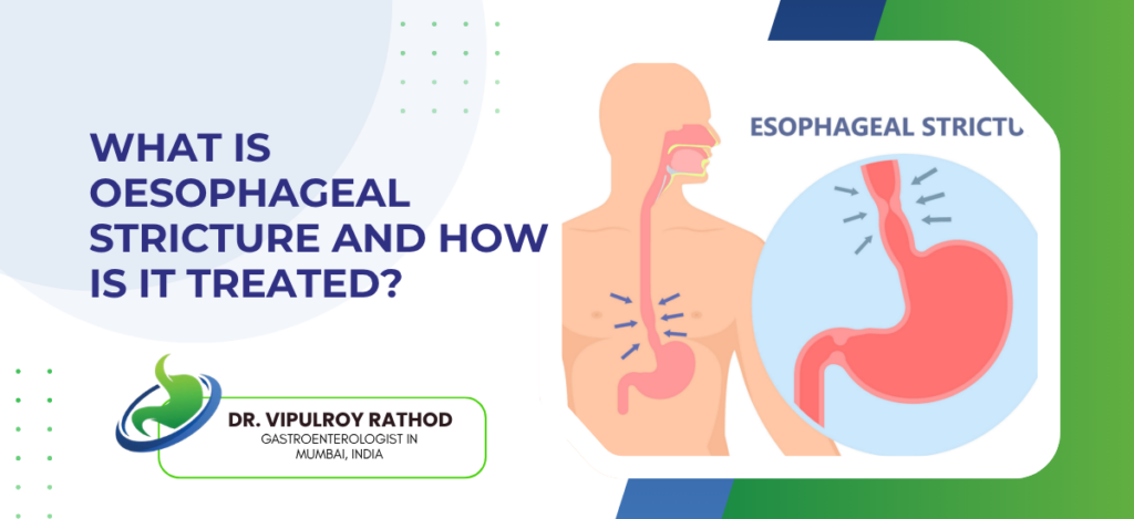
What Is Oesophageal Stricture and How Is It Treated?

Oesophageal stricture is narrowing of the oesophagus that obstructs the passage of food, caused most commonly by chronic acid reflux but also by caustic injury, radiation, eosinophilic oesophagitis, post-surgical scarring, or malignancy. The classic symptom is dysphagia, food sticking in the chest, usually solids before liquids, often progressive over months. Treatment is endoscopic in the majority of cases, with balloon dilatation or bougie dilatation reopening the narrowed segment. Underlying cause needs treatment alongside, because dilatation alone without addressing reflux or inflammation rarely produces lasting relief. According to Dr. Vipulroy Rathod, Gastroenterologist in Mumbai, “Strictures get treated successfully in most patients with endoscopic dilatation. The challenge isn’t the procedure. It’s stopping the stricture from coming back. PPIs for reflux strictures. Steroid injections for refractory cases. Treatment of underlying eosinophilic oesophagitis when that’s the cause. The dilatation opens the lumen. The maintenance keeps it open.” What causes oesophageal stricture? The causes fall into a few clear categories, and identifying the cause matters because it changes the treatment approach. Peptic stricture from chronic reflux. The commonest cause. Long-standing acid exposure damages the lower oesophagus, scarring develops, and the lumen narrows progressively over years. Usually occurs in patients with longstanding untreated or undertreated reflux. Eosinophilic oesophagitis produces strictures from chronic allergic inflammation. Typically affects younger patients and presents with food impaction or dysphagia. The strictures are often multiple and located higher in the oesophagus than peptic strictures. Caustic and radiation injury. Accidental or intentional ingestion of acid or alkali burns the oesophagus, with scarring developing weeks later. Radiation strictures occur after treatment for head, neck, or thoracic cancers and tend to be tight, long-segment, and harder to dilate. Malignant strictures from oesophageal or gastric cancer. These need biopsy before any dilatation. The treatment pathway shifts to oncology rather than continued endoscopic dilatation, with stenting as a palliative option in unresectable disease. The cause changes everything about management. Endoscopy in Mumbai services with biopsy capability during the same dilatation session avoid the need for repeat procedures and ensure cancer is ruled out at the earliest opportunity, particularly in older patients or those with weight loss alongside the dysphagia. How oesophageal stricture is treated? Treatment is mostly endoscopic. Surgery is reserved for the small number of cases where dilatation has failed repeatedly or the underlying cause is malignant and resectable. Balloon dilatation. A balloon catheter is passed through the scope to the stricture and inflated to a controlled diameter. Quick, well-tolerated, performed under sedation. Often the first-line endoscopic treatment for most peptic and inflammatory strictures. Bougie dilatation uses graduated dilators passed sequentially over a guidewire. Useful for longer strictures or those that don’t fully respond to balloon dilatation. Either technique works in expert hands. Steroid injection. Triamcinolone injected into the stricture site reduces scarring and prolongs the interval before re-stricture in refractory cases. Used selectively rather than universally, mostly in patients needing repeat dilatations within short intervals. Stenting for malignant or refractory benign strictures. Self-expanding metal stents reopen the lumen and stay in place for weeks to months, used mostly in palliative oncology cases or in benign strictures that have failed multiple dilatations. Treating the cause is what prevents recurrence. Read more on eosinophilic oesophagitis for one of the increasingly recognised causes of oesophageal stricture, particularly in younger patients who don’t fit the classical reflux pattern. Why choose Dr. Vipulroy Rathod for oesophageal stricture treatment? Dr. Vipulroy Rathod has been performing endoscopic dilatation and stricture management at Fortis Hospital Mulund for over three decades, with the technical experience across balloon and bougie techniques that complex strictures often require. Many patients arrive after multiple dilatations elsewhere that didn’t address the underlying cause, with the stricture returning every few months and the same dilatation being repeated as a recurrent procedure rather than a step in a coherent plan. The bigger picture in stricture management is whether the underlying disease is being treated alongside the mechanical narrowing. PPIs at proper doses for reflux strictures. Topical steroids for eosinophilic oesophagitis. Surveillance and biopsy in older patients to exclude malignancy. Dilatation alone, without that broader workup, is what produces patients who come back every three months for years. Book your consultation today with one of India’s most experienced specialists for oesophageal stricture assessment and endoscopic treatment. Book Appointment Call now Frequently Asked Questions Is endoscopic dilatation painful? No, the procedure is done under sedation. Mild throat discomfort for a day afterwards is normal. How long does relief from dilatation last? Months to years, depending on the cause and whether the underlying disease is being treated alongside. Can oesophageal stricture come back after treatment? Yes, particularly if the underlying cause isn’t controlled. Refractory strictures sometimes need repeated dilatations or steroid injections. When is surgery needed for stricture? Rarely. Mostly when dilatation has failed multiple times, when stricture is associated with resectable malignancy, or with complications like perforation. Reference links- Esophageal Dilation Guidelines, American Society for Gastrointestinal Endoscopy — https://www.asge.org/home/practice-support/guidelines Management of Esophageal Strictures, World Gastroenterology Organisation — https://www.worldgastroenterology.org/guidelines

What Is Oesophageal Stricture and How Is It Treated? Read More »

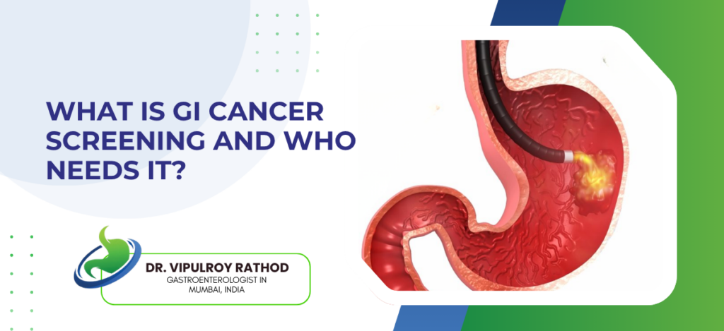
What Is GI Cancer Screening and Who Needs It?

GI cancer screening is the use of tests to find gastrointestinal cancers before symptoms develop. The cancers it targets are colorectal, oesophageal, gastric, pancreatic, and liver. The aim is early detection at a stage where treatment is curative rather than palliative. Colonoscopy remains the most effective screening test. Upper GI endoscopy, FibroScan, ultrasound, and EUS are used selectively depending on risk. Stage one GI cancers have survival above ninety percent. That number falls sharply at later stages. That’s the whole point of screening. According to Dr. Vipulroy Rathod, Gastroenterologist in Mumbai, “Most GI cancers stay silent. No early symptoms. By the time something appears, obstruction, bleeding, weight loss, the curative window has closed considerably. Screening exists to close that gap before the patient ever knows there’s one. The ones who screen do better. The ones who wait for symptoms usually arrive late.” Which are the main GI cancer screening tests and what they detect? Each test targets specific cancers. Choosing the right combination depends on the patient’s risk rather than applying a generic package. Colonoscopy. The single most effective screening test in GI medicine. Detects and removes precancerous polyps before they turn into cancer, and identifies colorectal cancer at stages where surgical cure is realistic. Upper GI endoscopy looks for oesophageal and gastric cancers along with Barrett’s oesophagus as a precursor. Used selectively, mostly in patients with reflux history or specific risk factors rather than universally. FibroScan and liver imaging. Identifies cirrhosis and high-risk metabolic liver disease. Cirrhotic patients then go on six-monthly ultrasound surveillance for hepatocellular carcinoma. EUS for pancreatic surveillance. Strong family history. Genetic syndromes. High-risk cystic lesions found on imaging. The screening plan should fit the patient, not the other way around. Endoscopy in Mumbai services that can combine colonoscopy, upper GI endoscopy, and liver assessment in a single visit make screening practical for working adults who can’t take repeated time off for separate appointments. Who needs GI cancer screening? Not every adult needs every test. Risk factors and family history decide which tests, at what age, and how often. Average-risk colorectal screening. Starts at 45 to 50 depending on guideline, repeated every ten years if results stay normal. Patients with polyps on previous scopes follow shorter intervals. Family history changes the schedule. First-degree relative with colorectal cancer. Screening starts ten years before that relative’s age at diagnosis, or at 40, whichever comes first. Cirrhosis and chronic liver disease. Six-monthly ultrasound plus alpha-fetoprotein for hepatocellular carcinoma surveillance, regardless of what caused the underlying liver disease. Hereditary GI cancer syndromes need specific protocols. Lynch syndrome, FAP, hereditary diffuse gastric cancer. Earlier start, more frequent intervals, often guided by genetics services. The benefit of screening depends on the right test reaching the right patient at the right time. Read more on digestive health screening after 50 for how the screening agenda evolves with age and risk factors. Why choose Dr. Vipulroy Rathod for GI cancer screening? Dr. Vipulroy Rathod has been doing GI cancer screening and surveillance work at Fortis Hospital Mulund since the late 1990s, with the kind of volume that builds pattern recognition for early cancers most clinicians never get a chance to develop. Screening plans get tailored to family history, metabolic risk, and previous findings rather than applied as a generic checkup package. The bigger problem patients face isn’t access. It’s whether the right tests get ordered in the right order. Some arrive after years of generic wellness packages that never included a colonoscopy. Others arrive having had multiple stool tests when family history warranted a scope from the start. Matching the test to the patient is what makes screening actually save lives. Book your consultation today with one of India’s most experienced specialists for GI cancer screening and surveillance. Book Appointment Call now Frequently Asked Questions At what age should GI cancer screening start? Average-risk colorectal screening begins at 45 to 50. Family history or specific risk factors mean starting earlier. Is screening necessary if I have no symptoms? Yes, especially then. How often should screening be repeated? Normal colonoscopy usually allows a ten-year gap. Polyps, family history, or other risk factors shorten the interval. Are blood tests enough for GI cancer screening? No. Tumour markers like CEA aren’t sensitive enough for screening, and colonoscopy and endoscopy remain the standard. Reference links- Colorectal Cancer Screening Guidelines, American College of Gastroenterology — https://gi.org/guideline/colorectal-cancer-screening/ GI Cancer Surveillance Standards, World Gastroenterology Organisation — https://www.worldgastroenterology.org/guidelines

What Is GI Cancer Screening and Who Needs It? Read More »

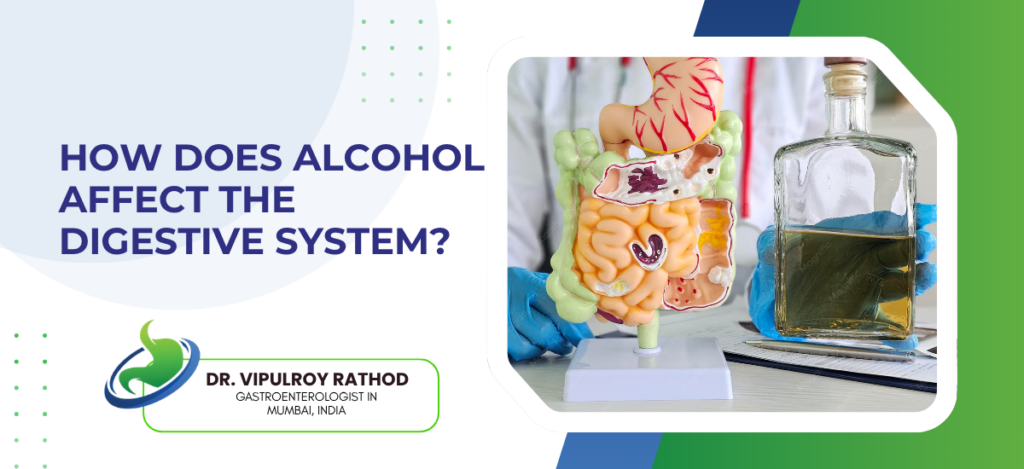
How Does Alcohol Affect the Digestive System?

Alcohol affects every part of the digestive system, from the oesophagus to the colon, with the liver and pancreas taking the most significant damage at higher intakes. It irritates the mucosal lining, alters gut motility, disrupts the microbiome, and at higher doses causes structural injury including fatty liver, alcoholic hepatitis, chronic pancreatitis, and gastrointestinal cancers. The damage is dose-related and partly genetic, with some patients showing significant disease at intakes others tolerate without obvious problems. According to Dr. Vipulroy Rathod, Gastroenterologist in Mumbai, “Patients often think alcohol only affects the liver. The truth is wider than that. The oesophagus, the stomach, the pancreas, the gut microbiome, all of them take damage. The liver gets attention because it’s the organ that fails most visibly. Many of the symptoms patients come in with, reflux that won’t settle, gastritis, recurrent diarrhoea, episodes of pancreatitis, trace back to the same drinking pattern that the patient hasn’t yet been asked about.” Where alcohol damages the digestive system? The injury patterns differ by organ. Some are direct mucosal effects, others come from systemic metabolic consequences. Oesophagus. Alcohol relaxes the lower oesophageal sphincter, worsening reflux. Long-term drinking raises the risk of Barrett’s oesophagus and squamous cell cancer, particularly in heavy drinkers who smoke. The stomach takes direct mucosal damage. Acute gastritis with heavy drinking, chronic gastritis with regular use, and a raised risk of peptic ulcer disease especially with NSAIDs. Pancreas. Alcohol is the commonest cause of chronic pancreatitis worldwide. The damage accumulates with each acute episode, leaving scar tissue that produces diabetes and enzyme insufficiency over years. Liver injury runs from fatty liver to alcoholic hepatitis to cirrhosis. The progression depends on dose, duration, and genetics. Fatty liver reverses with abstinence. Cirrhosis does not. The damage doesn’t always announce itself early. Endoscopy in Mumbai services with upper and lower GI assessment alongside liver elastography in the same workup help patients with significant alcohol use, because the asymptomatic phase of most alcohol-related disease lasts years. What changes when alcohol intake stops? The digestive system has substantial capacity for recovery if exposure stops early enough. The rate of reversal depends on which organ and how much damage has already accumulated. Stomach and oesophagus recover quickly. Reflux improves within weeks. Gastritis resolves over a similar period. Mucosal healing is usually complete within months. The pancreas is harder. Acute pancreatitis recovers over weeks, but the structural damage from chronic alcohol-related pancreatitis is permanent. Stopping prevents further injury and reduces flare frequency. Liver recovery depends on stage. Fatty liver reverses fully within three to six months. Alcoholic hepatitis can resolve but may leave fibrosis. Cirrhosis doesn’t reverse, though stopping extends survival meaningfully. The gut microbiome shifts back over months. Inflammation markers come down. Mood and metabolic parameters improve. The pattern of recovery here is consistent across recent research. Recovery isn’t uniform, and the staging dictates what’s realistic. Read more on fatty liver and metabolic health for how alcohol-related liver injury fits into the broader category of metabolic and toxic liver disease. Why choose Dr. Vipulroy Rathod for alcohol-related digestive disease? Dr. Vipulroy Rathod has been managing alcohol-related digestive disease at Fortis Hospital Mulund for over three decades, with experience across the spectrum from early reflux and gastritis to established chronic pancreatitis and decompensated liver disease. Many patients arrive after years of being treated symptomatically for one part of the picture, without anyone having asked about alcohol intake systematically or staged the overall damage properly. The conversation about alcohol intake is one most clinicians skip because it’s uncomfortable. The result is patients who have multiple organs being damaged by the same exposure, getting partial treatment for each symptom, without anyone connecting the dots. Asking properly and assessing each organ honestly is what makes effective treatment possible. Book your consultation today with one of India’s most experienced specialists for alcohol-related digestive disease and integrated GI assessment. Book Appointment Call now Frequently Asked Questions How much alcohol is safe for the digestive system? There is no completely safe threshold. Recent evidence shows even moderate drinking raises the risk of GI cancers and worsens metabolic liver disease. Can alcohol damage be reversed? Some of it. Fatty liver reverses fully with abstinence, gastritis and reflux resolve quickly, but chronic pancreatitis and cirrhosis don’t fully reverse. Is occasional binge drinking less harmful than regular drinking? Not really. Binge episodes are particularly damaging to the pancreas and can trigger acute pancreatitis on their own. Are there tests that show alcohol-related damage before symptoms appear? Yes. Liver function tests, FibroScan, faecal elastase, and endoscopy can identify problems years before clinical disease develops. Reference links- Alcohol-Related Liver Disease Guidelines, American Association for the Study of Liver Diseases — https://www.aasld.org/sites/default/files/2022-04/AASLD%20ALD%20Guidance%202019%20Update.pdf Alcohol and Gastrointestinal Health, World Gastroenterology Organisation — https://www.worldgastroenterology.org/guidelines

How Does Alcohol Affect the Digestive System? Read More »

What Is GI Bleeding and When Is It Serious?

Gastrointestinal bleeding is any bleeding from the digestive tract, anywhere from oesophagus to rectum. It becomes serious when the bleed is fast enough to cause haemodynamic changes, when haemoglobin drops significantly, when symptoms of hypovolaemia develop, or when the suspected source is a high-risk lesion like a peptic ulcer with a visible vessel or oesophageal varices. The location, the rate, and the underlying cause together determine urgency, and any one of those in isolation can underestimate or overestimate the situation. According to Dr. Vipulroy Rathod, Gastroenterologist in Mumbai, “Patients misjudge GI bleeding in both directions. Some panic over a streak of red blood from a haemorrhoid that’s never dangerous. Others ignore black tarry stools for weeks, which is much more concerning. Melena means significant upper GI bleeding. The colour matters. The volume matters. The symptoms matter. All three together tell the story, and any one in isolation can mislead.” What GI bleeding looks like clinically? GI bleeding presents differently depending on where it’s coming from and how fast it’s happening. Recognising the pattern is the first step in working out how urgent things are. Upper GI bleeding. Originates above the ligament of Treitz. Oesophagus, stomach, or duodenum. Presented as haematemesis, fresh red or coffee-ground vomitus. Or as melena, black tarry stools from digested blood passing through the small bowel. Occasionally as fresh red blood per rectum if the bleed is massive and rapid. Lower GI bleeding originates below the ligament, from the small bowel, colon, or rectum. It usually appears as haematochezia, fresh or maroon blood mixed with stool, sometimes with clots. Common causes include diverticular disease, haemorrhoids, anal fissures, colorectal cancer, and inflammatory bowel disease. Occult bleeding. Slow, ongoing blood loss that the patient never sees. Shows up as iron deficiency anemia on routine bloods, sometimes with a positive faecal occult blood test. Often the only sign of a small but persistently bleeding lesion that needs endoscopic investigation. Obscure GI bleeding is the term used when upper and lower endoscopy have both come back negative but bleeding continues. The small bowel is usually the source. Investigation needs capsule endoscopy, push enteroscopy, or CT angiography depending on whether the bleed is overt or obscure. The colour and pattern usually narrow the location down before any test is done. Advanced GI procedures then confirm the source and treat it in the same session in most cases, with endoscopic clips, cautery, band ligation, or injection therapy depending on the lesion. When GI bleeding becomes serious? Most GI bleeding episodes are minor and self-limiting. The ones that aren’t tend to declare themselves through specific clinical features. Haemodynamic compromise is the clearest marker. Tachycardia. Low blood pressure. Dizziness on standing. Sometimes frank shock. These signs reflect significant volume loss, often before the patient has appreciated how much bleeding has actually occurred. Resuscitation comes first in these cases, with diagnosis following once the patient is stable. Melena or haematemesis both indicate upper GI bleeding that has reached a volume significant enough to be obvious to the patient. Coffee-ground vomiting suggests slower or older bleeding, fresh red haematemesis suggests active brisk bleeding, and melena typically means at least 50 to 100 mL of blood has been lost into the upper tract. Bleeding in patients with known risk factors changes the threshold completely. Cirrhosis with portal hypertension raises the risk of variceal bleeding, which carries high mortality without urgent endoscopic intervention. Patients on anticoagulants, antiplatelet agents, or NSAIDs have higher bleed rates and worse outcomes when bleeding does occur. A first episode in any of these groups warrants admission regardless of how stable the patient looks initially. Recurrent bleeding matters even when individual episodes seem minor. Multiple small bleeds point to an underlying lesion that hasn’t been identified, and the cumulative effect on haemoglobin and quality of life justifies investigation rather than continuing to manage each episode separately. Read more on endoscopic treatment for GI bleeding for how modern endoscopic techniques manage most significant bleeds without surgical intervention. Why choose Dr. Vipulroy Rathod for GI bleeding assessment? Dr. Vipulroy Rathod has been managing acute and chronic GI bleeding at Fortis Hospital Mulund for over three decades, with the urgent endoscopy capability that serious cases need. Patients arrive at various stages. Some after a single concerning episode. Others after months of intermittent bleeding that’s been managed symptomatically without anyone identifying the source. Both groups need proper assessment, often with endoscopy as the next step rather than another round of empirical iron or PPI treatment. The judgment that matters most in GI bleeding is timing. Endoscopy too early in a stable patient with self-limiting bleeding adds risk without changing outcomes. Endoscopy too late in a patient with ongoing bleeding allows the haemoglobin to drop further and complications to develop. Getting that balance right is what experience teaches. Book your consultation today with one of India’s most experienced specialists for GI bleeding assessment and endoscopic management. Book Appointment Call now Frequently Asked Questions Is bright red blood from the rectum always serious? Not always. A small streak on toilet paper or surface of stool is usually haemorrhoids or an anal fissure. Heavier red bleeding mixed with stool, recurrent episodes, or any bleeding alongside weight loss, change in bowel habit, or anaemia needs investigation regardless of how it looks. What does black tarry stool mean? Melena. Which is significant upper GI bleeding. The black colour comes from digested blood that has passed through the small intestine. Usually means at least 50 to 100 mL has been lost into the upper tract, and it shouldn’t be managed at home. Can GI bleeding stop on its own? Yes, many episodes do. Self-limiting bleeds from small ulcers, haemorrhoids, or minor inflammation often resolve without intervention. The question isn’t only whether it stops, but whether the source has been identified, because recurrence is common when the underlying lesion remains untreated. When should I go to the emergency department? If you’re vomiting blood. If your stools are black and tarry. If there’s heavy red rectal

What Is GI Bleeding and When Is It Serious? Read More »

What causes chronic diarrhoea and when to see a specialist? header with a person clutching their abdomen near a bathroom illustration (informational banner).

What Causes Chronic Diarrhoea and When to See a Specialist?

Chronic diarrhoea, defined as loose stools lasting more than four weeks, is caused most commonly by irritable bowel syndrome, inflammatory bowel disease, malabsorption syndromes, or chronic infections, with rarer causes including microscopic colitis, bile acid malabsorption, and gastrointestinal malignancy. Specialist referral is warranted when alarm features are present, when symptoms persist despite first-line treatment, or in patients over fifty regardless of severity. The symptoms overlap considerably between these conditions, and a structured workup is what separates them. According to Dr. Vipulroy Rathod, Gastroenterologist in Mumbai, “Most chronic diarrhoea in Indian patients gets treated empirically with antibiotics or anti-motility agents without anyone working out what’s actually going on. Some have IBS and need lifestyle advice. Some have IBD that’s been missed for years. Some have lactose intolerance or coeliac disease that a simple test would have picked up. Empirical treatment without diagnosis is how the underlying cause stays hidden.” What are the common causes of chronic diarrhoea? The diagnosis usually narrows down based on stool pattern, associated symptoms, and a few targeted tests rather than a shotgun approach. Irritable bowel syndrome. The commonest cause overall. Crampy pain, alternating bowel habits in some patients, urgency, no blood, no weight loss. IBS is a real diagnosis with biological underpinnings, not a label for unexplained symptoms. It is also a diagnosis of exclusion that requires ruling out organic causes first. Inflammatory bowel disease produces diarrhoea with blood, urgency, nocturnal symptoms, weight loss, and raised inflammatory markers. Patients often spend years being treated for IBS before someone runs a calprotectin or arranges a colonoscopy. Malabsorption syndromes. Coeliac disease, pancreatic insufficiency, bile acid malabsorption, small intestinal bacterial overgrowth. These cause bulky, fatty, sometimes pale stools, often with weight loss and bloating disproportionate to the diarrhoea. Testing for each is straightforward once the pattern is recognised. Microscopic colitis often gets missed because the colon looks normal on endoscopy. Diagnosis requires biopsies even when the mucosa appears unremarkable. The condition affects older patients particularly and is sometimes triggered by NSAIDs, PPIs, or SSRIs. A few less common but important causes need to stay on the radar. Endoscopy in Mumbai services with colonoscopy, random biopsy capability, push enteroscopy, and capsule endoscopy in selected cases are what allow these diagnoses to be made when the standard workup hasn’t given clear answers. When chronic diarrhoea warrants specialist referral? Not every patient with chronic diarrhoea needs a gastroenterologist. Specific features should prompt referral rather than another round of empirical treatment. Alarm symptoms come first on the list. Blood in the stool. Weight loss. Nocturnal diarrhoea waking the patient from sleep. Iron deficiency anaemia. Family history of colorectal cancer or IBD. Any one of these features changes the workup completely, and they shouldn’t be managed in primary care without specialist input. Age matters as much as symptoms. Chronic diarrhoea starting after fifty has a different differential than the same symptoms in a twenty-five-year-old. The probability of inflammatory bowel disease falls with age, while microscopic colitis, ischaemic colitis, and malignancy rise. The threshold for colonoscopy is lower in this group regardless of how mild the symptoms appear. Failure to respond to first-line management is another clear trigger. Patients tried on dietary modification, anti-motility agents, or empirical antibiotics for several weeks without improvement need a proper workup. Persisting with treatments that aren’t working delays the actual diagnosis. The fourth situation is when the diagnosis matters for the patient’s long-term care. Coeliac disease needs lifelong gluten avoidance. IBD needs specific treatment and surveillance. Bile acid malabsorption responds to cholestyramine. None of these can be managed properly without a diagnosis, and trial-and-error rarely arrives at the right answer. Read more on what IBD is and how it’s diagnosed for one of the more important differential diagnoses in chronic diarrhoea that frequently gets missed in primary care. Why choose Dr. Vipulroy Rathod for chronic diarrhoea assessment? Dr. Vipulroy Rathod has been assessing and managing chronic diarrhoea at Fortis Hospital Mulund for over three decades, with the structured workup that separates IBS from IBD from malabsorption from rarer diagnoses without putting every patient through every test. The history and examination drive most of what gets tested, but the threshold for organic disease screening is appropriately low in patients with any alarm features or with a presentation that doesn’t fit cleanly into IBS. The bigger problem patients face isn’t access to tests. It’s getting the right tests in the right order. Some patients arrive having had multiple stool cultures and ultrasounds when a colonoscopy with biopsies would have answered the question in one appointment. Others arrive after years of being managed for IBS when their actual diagnosis was coeliac disease or microscopic colitis. Working through the differential properly is what makes the difference. Book your consultation today with one of India’s most experienced specialists for chronic diarrhoea assessment and diagnosis. Book Appointment Call now Frequently Asked Questions When is diarrhoea considered chronic? When it lasts more than four weeks. Anything shorter is usually acute or subacute, with a different differential weighted heavily towards infection. Should every patient with chronic diarrhoea have a colonoscopy? No. The threshold depends on age, alarm features, and clinical pattern. Young patients with classical IBS features and no alarm symptoms often don’t need a scope. Older patients, those with any alarm feature, or those who haven’t responded to first-line management usually do. What blood and stool tests are usually done? Full blood count, CRP, faecal calprotectin, stool infection screen, coeliac serology, and thyroid function as the starting set. Pancreatic faecal elastase, bile acid testing, and SIBO breath tests come in selectively based on the clinical picture. Can chronic diarrhoea resolve on its own? Yes, in functional cases like post-infectious IBS that improves over months. Persistent organic disease doesn’t usually resolve without specific treatment, which is why a proper diagnosis matters before assuming things will settle. Reference links- Chronic Diarrhoea Guidelines, American College of Gastroenterology — https://gi.org/guideline/diarrhea-in-the-adult/ Chronic Diarrhoea Management Standards, World Gastroenterology Organisation — https://www.worldgastroenterology.org/guidelines

What Causes Chronic Diarrhoea and When to See a Specialist? Read More »

IBD Flare Management: What to Do When Symptoms Return?

A flare is the return of active IBD symptoms after a period of remission. Patients usually know what their flare looks like, because the symptoms tend to follow the same pattern that brought them to a doctor in the first place. Stool frequency rises, blood may reappear, abdominal pain creeps back, fatigue and weight loss follow if the flare runs long enough. The instinct to wait it out is one of the commoner mistakes patients make. Acting early shortens flares and prevents the damage that’s much harder to undo later. According to Dr. Vipulroy Rathod, Gastroenterologist in Mumbai, “Patients wait too long. They hope it’ll settle on its own. Sometimes it does. Often it doesn’t, and what could have been managed with a steroid course or a biologic dose tweak in week one ends up needing hospitalisation by week four. Acting early when it turns out to be nothing costs much less than acting late when it turns out to be a major flare.” What to do when symptoms return? The first response isn’t a Google search. It’s a structured assessment, ideally with input from the gastroenterology team within days, not weeks. Document symptoms specifically. Stool frequency per day. Presence and amount of blood. Pain pattern. Weight change. Vague reports like “things are worse” don’t give the clinical team enough to work with. Check medication adherence honestly. Missed doses, stopped biologics, irregular maintenance therapy, these are the commonest preventable causes of flare. Patients are often reluctant to admit gaps, but the team needs to know because management changes depending on whether the flare comes from missed treatment or genuine breakthrough disease. Always rule out infection before escalating immunosuppression. Stool testing for C. difficile, bacterial pathogens, and parasites is non-negotiable before adding steroids. Giving steroids to a patient with superimposed C. difficile colitis makes things significantly worse. Contact the gastroenterology team early. Many flares can be assessed remotely at first, with stool tests, blood markers, and history guiding the next step. IBD treatment plans should include clear flare instructions agreed in advance, telling patients which symptoms warrant immediate contact, what tests will be ordered, and what the escalation threshold looks like, so the response is structured rather than reactive. What clinical assessment and escalation actually involves? Once a flare is reported, the workup follows a familiar pattern. Confirm active inflammation. Rule out other causes. Decide how aggressively to escalate. Stool tests come first because they answer the most important question quickly. Faecal calprotectin to quantify inflammation. Cultures and PCR for infection. Calprotectin above 250 in a patient with returned symptoms strongly suggests active disease. Under 100 raises the possibility that something else is going on entirely, sometimes IBS-like symptoms in patients with healed bowel, sometimes a non-IBD cause that’s been missed. Blood tests cover inflammatory markers (CRP and ESR), full blood count, liver function, and a basic metabolic panel. Anaemia, low albumin, raised CRP all support active disease. In biologic patients drug levels go in at the same time, because the commonest reason for biologic failure isn’t drug resistance, it’s underdosing. Imaging or endoscopy isn’t always needed at first contact. Mild to moderate flares are often managed on stool and blood tests alone in the early phase. Severe symptoms, suspected complications, or flares not responding to first-line treatment usually trigger CT enterography, MR enterography, or sigmoidoscopy depending on availability and what’s clinically most useful. Treatment escalation runs on a ladder rather than a single decision. Optimise maintenance therapy. Add a short course of steroids if needed for symptom control. Consider biologic dose escalation or switching agents if the flare reflects genuine loss of response. Surgery for cases that don’t respond to medical rescue, or where complications have developed. Read more on when IBD needs surgery for how the surgical option fits into flare management when medical treatment isn’t enough. Why choose Dr. Vipulroy Rathod for IBD flare management? Dr. Vipulroy Rathod has been managing IBD flares and the longer trajectory of inflammatory bowel disease at Fortis Hospital Mulund for over three decades, with the kind of structured flare protocols that come from seeing the same patterns repeatedly and knowing which escalation works for which patient. Many patients arrive mid-flare having waited too long, when earlier intervention would have made the difference between outpatient management and a hospital admission. The pattern that matters most in flare management is recognition. Patients who know what their flare looks like, who have a clear plan agreed with their team, who contact the clinic at day three rather than week three, consistently do better than patients trying to manage flares alone. The work upfront on agreeing what to do when symptoms return pays back many times over. Book your consultation today with one of India’s most experienced specialists for IBD assessment and structured flare management. Book Appointment Call now Frequently Asked Questions How quickly should I contact the clinic during a flare? Within a few days. Don’t wait for symptoms to become severe. Early assessment gives the team more options than late assessment. Can I just take more of my current medication? Sometimes, often not. Some flares respond to maintenance optimisation. Others need short steroid courses or biologic adjustment. Self-adjusting without team input risks under-treating or masking infection. What blood and stool tests are usually done? Faecal calprotectin and stool infection screen first. Then CRP, full blood count, liver function. Biologic drug levels if applicable. Most results come back within a few days. Will I need hospital admission? Most flares are managed as outpatients. Admission is reserved for severe colitis not responding to oral treatment, complications like obstruction or perforation, or patients too unwell to manage at home. Reference links- IBD Flare Management Guidelines, American Gastroenterological Association — https://gastro.org/clinical-guidance/ ECCO Topical Review on IBD Flare, European Crohn’s and Colitis Organisation — https://www.ecco-ibd.eu/publications/ecco-guidelines.html

IBD Flare Management: What to Do When Symptoms Return? Read More »

Scroll to Top
Call Now Button