 Skip to content
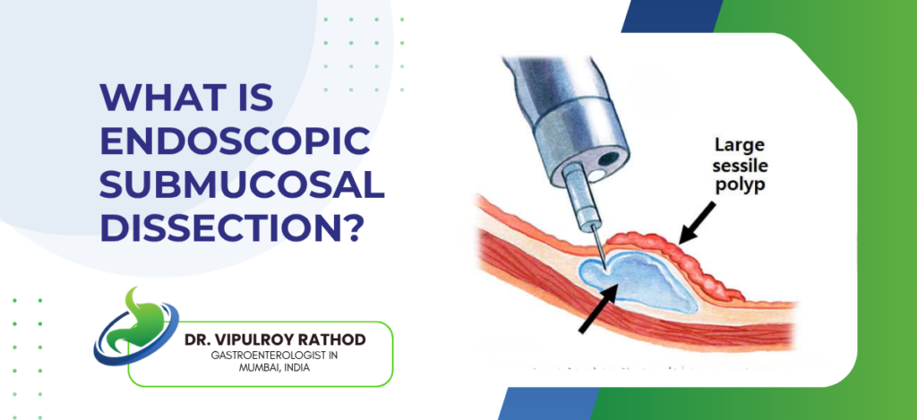
Endoscopic submucosal dissection (ESD) is an advanced endoscopic technique that removes early-stage GI cancers, large polyps, and submucosal lesions in a single intact piece through the scope. The procedure uses a specialised electrosurgical knife passed through a standard endoscope, allowing en bloc resection of lesions up to several centimetres in size without open or laparoscopic surgery.
According to Dr. Vipulroy Rathod, Gastroenterologist in Mumbai, “ESD is what changed the management of early GI cancer. Lesions that needed gastrectomy or oesophagectomy can now be removed through a scope, in one piece, with the patient going home the next day. The key is en bloc resection. That gives the pathologist a complete specimen and tells us whether the cancer was confined to the mucosa or has gone deeper, which decides whether anything else needs to follow.”
What conditions are treated with ESD?
ESD is reserved for lesions where en bloc resection is essential, mainly early cancers and large or complex polyps.
- Early gastric cancer. Tumours confined to the mucosa or superficial submucosa, without lymph node involvement, are the classic ESD indication. Cure rates match surgical resection in selected cases.
- Early oesophageal cancer including squamous cell carcinoma and Barrett’s adenocarcinoma in superficial stages. ESD provides definitive treatment while preserving the oesophagus completely.
- Large colorectal polyps and laterally spreading tumours. Lesions over 20 mm or those with features suggesting they can’t be safely removed by piecemeal EMR. ESD allows complete en bloc removal regardless of size.
- Submucosal tumours and certain neuroendocrine tumours in selected locations and sizes. ESD or tunnelling techniques remove these without breaching the muscle layer or needing surgery.
The decision to do ESD depends on pre-procedure imaging assessment. Advanced endoscopy units with chromoendoscopy and EUS for accurate depth assessment select the right cases for ESD, distinguishing them from the lesions that need surgery instead.
How is ESD performed and what is recovery like?
The procedure uses a specialised electrosurgical knife and follows a structured sequence of marking, injection, incision, and dissection.
- Marking the margins. The borders of the lesion are marked with electrocautery dots, usually with a five-millimetre margin around visible disease. This defines the resection field.
- A submucosal injection lifts the lesion away from the muscle layer underneath, creating a working space. The solution is often mixed with dye to make the submucosal layer easier to see during dissection.
- Circumferential incision and dissection. The mucosa is incised along the marked margin, then the submucosal layer is dissected away from the muscle using the knife, freeing the lesion in one piece.
- Mucosal defect closure with clips at the end of the procedure. Most patients go home within twenty-four to forty-eight hours with normal activity resumed in a few days.
ESD has a learning curve and complication rates fall sharply with operator experience. Read more on endoscopic procedures for related advanced submucosal techniques used in different clinical situations.
Why choose Dr. Vipulroy Rathod for ESD?
Dr. Vipulroy Rathod has been performing advanced endoscopic resection techniques at Fortis Hospital Mulund for over three decades, including ESD for early GI cancers and large lesions that would otherwise require surgical resection. The training and equipment requirements for ESD are significant, and the procedure is performed at only a small number of centres in India with the volume needed to do it safely.
The reason ESD needs experienced hands is the dissection itself. The plane between the submucosa and muscle is narrow and the consequences of going wrong are serious. Perforation, bleeding, incomplete resection. The complication rate falls dramatically with operator experience, which is why ESD volume per operator matters more than equipment availability in any centre.
Book your consultation today with one of India’s most experienced specialists for ESD and advanced endoscopic resection.
Frequently Asked Questions
Yes, in appropriately selected cases. Cure rates match surgical resection when the lesion is correctly staged and the resection achieves clear margins.
Anywhere from one to four hours depending on the size and location of the lesion. Large gastric ESDs and complex colorectal lesions take longer than smaller superficial cases.
Bleeding, perforation, and incomplete resection are the principal complications. All three are uncommon in experienced hands and most can be managed endoscopically when they occur.
Limited. Only a small number of centres offer ESD with the equipment and volume needed for safe practice. Most patients are referred to high-volume specialist units.
Disclaimer:
This blog is intended for informational and educational purposes only and should not be considered medical advice.
Please consult a qualified gastroenterologist or healthcare professional for personalized diagnosis and treatment.
