 Skip to content
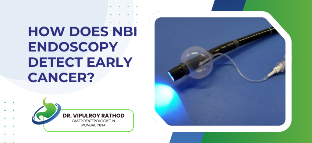
Narrow band imaging (NBI) is an advanced endoscopic technique that uses filtered blue and green light to highlight mucosal vessel and surface patterns, allowing detection of early cancers and precancerous lesions that standard white light endoscopy can miss. The technology is built into modern endoscopes and is switched on at the touch of a button during routine procedures.
According to Dr. Vipulroy Rathod, Gastroenterologist in Mumbai, “NBI changed how we look at the GI tract. The same scope, the same patient, but the lesions that white light barely shows light up clearly under NBI. Subtle Barrett’s dysplasia. Early gastric cancer in a chronic gastritis background. Flat colonic adenomas that would have been missed. It doesn’t replace careful inspection, but it makes careful inspection much more productive.”
What does NBI show and how does it work?
NBI works on the principle that haemoglobin absorbs specific wavelengths of light, which makes blood vessels appear with high contrast against surrounding tissue.
- Mucosal vessel patterns. NBI highlights the superficial capillary network on the mucosal surface. Early cancers produce irregular, distorted, or dense vessel patterns that stand out clearly from normal background mucosa.
- The pit pattern of glandular structures becomes more visible under NBI, which helps distinguish neoplastic from non-neoplastic lesions in the colon and stomach. Classifications like the Kudo and NICE systems are based on these patterns.
- Demarcation lines around lesions. NBI shows sharp borders between abnormal and normal mucosa more clearly than white light, which helps in deciding where the lesion ends and where the resection margin should be placed.
- Subtle colour changes from normal pink mucosa to brownish or whitish areas are amplified under NBI, particularly in the oesophagus where Barrett’s dysplasia and squamous neoplasia present as colour shifts before forming a visible mass.
NBI is most useful when combined with high-definition endoscopy and magnification. Endoscopy services using modern scopes with NBI capability allow careful targeted inspection of high-risk areas rather than relying on random biopsies alone, which is the older approach to screening for early disease.
Where is NBI most useful clinically?
NBI has the strongest evidence base in a few specific clinical situations where early cancer detection changes management.
- Barrett’s oesophagus surveillance. NBI improves detection of dysplasia in Barrett’s patients, allowing targeted biopsies of visible lesions rather than random four-quadrant biopsies of normal-looking mucosa.
- Early gastric cancer screening in patients with chronic atrophic gastritis or intestinal metaplasia. NBI with magnification picks up subtle lesions that look unremarkable under white light.
- Colorectal polyp characterisation. NBI helps distinguish hyperplastic from adenomatous polyps in real time, reducing unnecessary polypectomy and allowing optical diagnosis in selected cases.
- Squamous cell oesophageal cancer in high-risk patients, particularly heavy smokers and drinkers. NBI lights up flat brownish areas that suggest early squamous neoplasia.
NBI improves detection but it doesn’t replace endoscopic experience and time spent inspecting. Read more on Colon cancer screening for how NBI fits into the broader screening strategy across different GI cancer types.
Why choose Dr. Vipulroy Rathod for NBI endoscopy?
Dr. Vipulroy Rathod has been performing advanced endoscopy at Fortis Hospital Mulund for over three decades, with modern NBI-capable systems and the volume of cases that builds the pattern recognition NBI requires to be useful. The technology only adds value when the operator knows what they’re looking for, which is why NBI training matters as much as equipment availability in any centre.
The mistake some centres make is assuming NBI alone improves cancer detection. It doesn’t. NBI improves detection when used by experienced operators who do careful targeted inspection. In less experienced hands, the additional information gets overlooked or misinterpreted, which is part of why NBI uptake has been slower than expected in many countries despite the supporting evidence.
Book your consultation today with one of India’s most experienced specialists for NBI endoscopy and early GI cancer detection.
Frequently Asked Questions
Part of regular endoscopy. NBI is built into modern scopes and switched on during the procedure at the operator’s discretion.
No. The preparation for NBI endoscopy is the same as for any standard upper GI endoscopy or colonoscopy.
In selected cases yes, particularly for small colorectal polyps where optical diagnosis is supported by current guidelines. For most other situations, biopsy remains the standard for tissue confirmation.
Increasingly available in larger centres and tertiary hospitals, though not yet universal. Patients should ask whether the centre uses NBI-capable scopes for surveillance and high-risk screening procedures.
Disclaimer:
This blog is intended for informational and educational purposes only and should not be considered medical advice.
Please consult a qualified gastroenterologist or healthcare professional for personalized diagnosis and treatment.
