 Skip to content
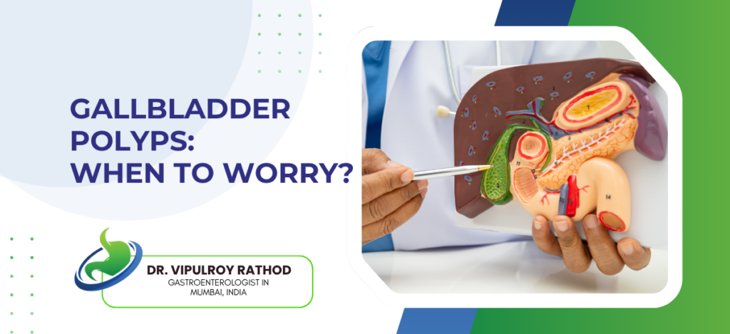
Gallbladder polyps are growths on the inner wall of the gallbladder, mostly benign and usually found incidentally on ultrasound. The concern is the small minority that can turn malignant or already contain cancer at the time of detection. Risk depends on size, age, presence of gallstones, and underlying conditions. Polyps under 10 mm in a young patient without other features rarely need anything beyond follow-up scans. Larger polyps, rapidly growing ones, or polyps in higher-risk patients usually need cholecystectomy. The judgment is in distinguishing which is which.
According to Dr. Vipulroy Rathod, Gastroenterologist in Mumbai, “Most gallbladder polyps are benign. Cholesterol polyps. Inflammatory polyps. Hyperplasia. None of those turn into cancer. So why do we still recommend follow-up for every polyp found on a routine scan? Because the small percentage that are adenomatous look identical on imaging, and the only way to know which group a patient is in is serial monitoring with surgical referral for the ones that show concerning features.”
What types of gallbladder polyps exist and which are concerning?
Polyps fall into a few categories. Most are non-neoplastic and stay benign throughout life.
- Cholesterol polyps. The commonest type. Around sixty percent of gallbladder polyps fall into this category, made of cholesterol-laden cells, small and often multiple. No malignant potential and rarely cause symptoms.
- Are inflammatory polyps dangerous? Almost never. These form after chronic gallbladder inflammation, usually alongside gallstones or recurrent cholecystitis, and have no cancer risk in themselves.
- Adenomatous polyps. True neoplastic lesions with malignant potential. Rare but the ones that drive surgical decisions. Risk of malignancy rises sharply with size, particularly above 10 mm.
- Adenomyomatosis is wall thickening that mimics a polyp on ultrasound. Not actually a polyp but often picked up the same way. Benign in itself, with imaging confirmation needed to distinguish it from a true polyp.
The features that change the workup are well-defined. Endoscopic ultrasound gives better resolution than transabdominal ultrasound for assessing polyp characteristics, particularly in lesions where the type isn’t clear and the size sits close to the surgical threshold.
When should a gallbladder polyp be removed?
The decision rests on a combination of size, growth, patient factors, and concerning imaging features rather than any single criterion.
- Size above 10 mm. The single most important risk factor. Polyps at this size carry significantly higher malignancy risk and most guidelines recommend cholecystectomy regardless of other factors.
- Growth on serial scans matters even in smaller polyps. A 6 mm polyp growing to 9 mm over a year often warrants surgery even though neither size alone would trigger the recommendation.
- Age above 50. Polyps in older patients carry higher malignant potential than equivalent-sized polyps in younger patients. The combined size and age threshold drops in this group.
- Other concerning features. Solitary polyp with broad base. Sessile rather than pedunculated morphology. Primary sclerosing cholangitis. Indian or East Asian ethnicity. Coexisting gallstones. Any of these raise the threshold for intervention.
Most patients don’t meet criteria and simply need follow-up scans. Read more on gallstone disease management for how gallbladder pathology fits into the broader picture, particularly when polyps and stones coexist.
Why choose Dr. Vipulroy Rathod for gallbladder polyp assessment?
Dr. Vipulroy Rathod has been managing biliary disease at Fortis Hospital Mulund since the late 1990s. Routine cholecystectomy cases. Complex polyp risk stratification. EUS-based assessment of indeterminate lesions. The patients who arrive after being told their polyp warrants urgent surgery without proper risk stratification. The patients who arrive having been told it’s nothing when family history and ethnicity put them in a higher-risk category. Both groups end up in the same clinic with very different management plans.
The judgment in gallbladder polyp management sits between two errors. Over-treat a benign cholesterol polyp and the patient gets unnecessary surgery. Under-treat a small adenomatous polyp that turns malignant over the following year and the consequences are far worse. Volume and proper imaging assessment are what keep both errors rare.
Book your consultation today with one of India’s most experienced specialists for gallbladder polyp assessment and surveillance.
Frequently Asked Questions
Rare cases of gallbladder polyps are cancerous.
Polyps under 6 mm need ultrasound every six to twelve months for two to three years. After that, less frequently if size and morphology stay stable, and the surveillance can be relaxed further or stopped completely in low-risk patients with consistently unchanged scans over a longer follow-up period.
Yes, Indian and East Asian patients carry higher malignant potential for the same polyp size.
Laparoscopic cholecystectomy is done for gallbladder polyps
Disclaimer:
This blog is intended for informational and educational purposes only and should not be considered medical advice.
Please consult a qualified gastroenterologist or healthcare professional for personalized diagnosis and treatment.
