Dr. Vipulroy Rathod

Author name: Dr. Rathod Medical Foundation

Bile Leakage After Gallbladder Surgery: Causes, Symptoms, and Treatment

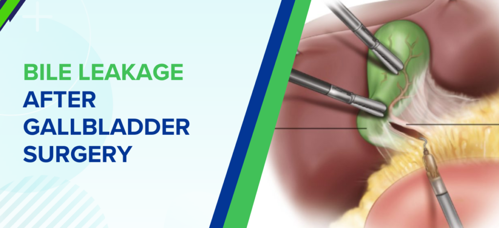
Gallbladder surgery, also known as cholecystectomy, is a widely performed procedure to treat gallstones, inflammation, and related digestive issues. It is generally safe and helps restore normal digestion by removing the diseased gallbladder. However, like any surgical procedure, certain complications may occur during the healing phase. One such complication is bile leakage after gallbladder surgery, where bile escapes from the bile ducts into the abdominal cavity instead of flowing into the intestine. This can lead to pain, infection, and delayed recovery if not addressed promptly. Dr. Vipulroy Rathod,a highly respected gastroenterologist in Mumbai, India, highlights, “Early identification of bile leakage is essential to prevent complications and ensure smooth healing.” He further adds, “With advanced endoscopic techniques, most cases can be managed effectively without the need for major surgery.” Dr. Rathod is widely recognized for his precision in managing complex biliary and pancreatic conditions. With extensive experience in advanced endoscopic procedures such as ERCP and EUS, he focuses on resolving post-surgical complications with minimally invasive techniques. His approach to managing bile leakage after gallbladder surgery emphasizes early intervention, accurate diagnosis, and targeted treatment, helping patients recover efficiently while minimizing the need for repeat procedures or prolonged hospitalization. Why does bile leakage happen after gallbladder removal? Let’s explore the common reasons behind this condition. What Causes Bile Leakage After Gallbladder Surgery? Bile leakage typically occurs when there is disruption or injury to the bile ducts during or after cholecystectomy. While it is not very common, certain factors can increase the risk. Common causes include:        Injury to bile ducts:Accidental damage during surgery can lead to leakage.        Cystic duct stump leak:Improper sealing of the duct after gallbladder removal may cause bile to escape.        Accessory bile ducts:Small, unnoticed ducts can leak bile if not identified during surgery.        Inflammation or infection:Pre-existing inflammation can weaken duct structures.        Surgical complications:Complex procedures may increase the risk of bile duct disruption. Understanding these causes helps in early detection and timely bile leakage treatment. Experiencing discomfort after surgery? Connect with a specialist to determine the underlying cause and next steps. Book Appointment Call now What signs should you watch for after surgery? Let’s discuss the symptoms that may indicate bile leakage. Common Symptoms of Bile Leakage After Gallbladder Surgery Symptoms of bile leakage can vary depending on the severity, but early warning signs should never be ignored. Common symptoms include:        Persistent abdominal pain, especially in the upper abdomen        Fever or chills, indicating possible infection        Nausea and vomiting        Swelling or bloating in the abdomen        Jaundice (yellowing of skin and eyes)        Drainage of bile fluid from surgical wounds (in some cases) “Recognizing these symptoms early can prevent complications and ensure faster recovery,” states Dr. Vipulroy Rathod. Now, let’s discuss the typical timeline of bile leakage after surgery. When Does Bile Leakage Typically Occur After Surgery? Bile leakage usually develops within the first few days after cholecystectomy, but in some cases, symptoms may appear later. Typical timeline:        Within 2–5 days:Most common period for symptoms to begin        Within 1–2 weeks:Delayed symptoms may occur in some patients        Rare cases:Late presentation if leakage is mild and gradual Early follow-up after surgery is important to detect any complications during this critical period. Let’s explore the diagnostic methods used to identify this condition. How is Bile Leakage Diagnosed? Accurate diagnosis is essential for effective bile leakage treatment and involves a combination of clinical evaluation and imaging. Diagnostic methods include: Ultrasound:Helps detect fluid accumulation in the abdomen CT scan:Provides detailed imaging of bile leakage and surrounding structures HIDA scan:Tracks bile flow to identify leaks MRCP (MRI scan):Visualizes bile ducts without invasive procedures ERCP (Endoscopic Retrograde Cholangiopancreatography):Both diagnostic and therapeutic These tests help locate the leak and guide appropriate treatment. How is bile leakage treated effectively? Let’s explore the treatment approaches available. Treatment Options for Bile Leakage After Gallbladder Surgery Treatment depends on the severity and source of the leak, with most cases managed using minimally invasive techniques. The treatment options include: Endoscopic Treatment (ERCP) This is the most commonly used approach, where an endoscope is used to access the bile duct and place a stent to redirect bile flow and reduce leakage. Biliary Stenting A small tube is inserted into the bile duct to keep it open and allow bile to flow correctly into the intestine, helping the leak to heal naturally. Sphincterotomy A minor procedure is done during ERCP in which a small cut is made to ease bile flow and reduce the pressure within the bile ducts. Percutaneous Drainage In case bile has built up in the abdomen, a drain may be placed through the skin to clear the fluid and prevent infection. Antibiotic Therapy Antibiotics are prescribed to treat or prevent infections caused by bile leakage. Surgical Repair (Rare Cases) Surgery can be performed to repair the bile duct in critical or complicated cases where minimally invasive intervention is not effective. Supportive Care and Monitoring Regular checkups, imaging and monitoring will be done to make sure that the leak is healing well and no further complications develop. Early and appropriate bile leakage treatment significantly improves recovery and reduces the risk of complications. What happens if bile leakage is ignored? Let’s understand the potential risks Complications if Bile Leakage is Left Untreated Untreated bile leakage can lead to serious complications, making early treatment essential. Possible complications include: Infection or abscess formation Peritonitis (inflammation of abdominal lining) Sepsis, a severe systemic infection Persistent abdominal pain and discomfort Delayed recovery and prolonged hospitalization Timely intervention can prevent these complications and improve outcomes. Worried about complications? Get in touch with a professional to avoid serious health risks and ensure smooth healing and long-term wellness. Book Appointment Call now What does recovery look like after treatment? Let’s explore the healing process Recovery and Healing After Bile Leak Treatment Recovery after bile leakage treatment depends on the severity of the condition and the type of treatment performed. Recovery highlights: Most patients recover well with minimally invasive procedures Symptoms improve within a few days after treatment Regular follow-up ensures proper healing Temporary stents may be removed

Bile Leakage After Gallbladder Surgery: Causes, Symptoms, and Treatment Read More »
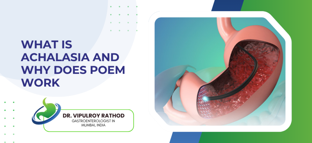
What Is Achalasia And Why Does POEM Work

Achalasia is a rare oesophageal motility disorder where the lower oesophageal sphincter fails to relax during swallowing and the oesophagus loses its coordinated muscle contractions, causing progressive difficulty swallowing both solids and liquids, regurgitation of undigested food, chest pain, and significant weight loss over time, POEM (Per-Oral Endoscopic Myotomy) works because it cuts the dysfunctional sphincter muscle from inside the oesophageal wall without any external incision, directly resolving the obstruction that medication and dilation can only temporarily manage. According to Dr. Vipulroy Rathod, Gastroenterologist in Mumbai, “Achalasia is one of those conditions where patients spend years being treated for reflux or anxiety before anyone thinks to investigate the oesophagus properly, and POEM changed the treatment landscape because it addresses the actual mechanical problem rather than working around it.” What Is Achalasia and How Does It Develop? Damaged nerve cells in the oesophageal wall are the root cause and once those cells are gone they don’t regenerate, which is why achalasia is managed not cured and why the intervention needs to be durable rather than temporary. Mechanism: Myenteric plexus neurons that coordinate lower oesophageal sphincter relaxation are progressively destroyed, likely through autoimmune processes in most cases, leaving a sphincter that stays contracted during swallowing and an oesophagus that can’t move food downward through organised peristalsis. Symptoms: Progressive dysphagia for both solids and liquids is the hallmark, regurgitation of undigested food hours after eating, chest pain from oesophageal spasm, and weight loss that gets attributed to anxiety or functional disorders for months before anyone orders a proper motility study. Diagnosis: High-resolution manometry is the gold standard showing absent peristalsis and incomplete LES relaxation, barium swallow shows the classic bird-beak narrowing at the gastro-oesophageal junction, endoscopy rules out malignancy causing pseudoachalasia which has identical symptoms but a completely different clinical picture. Types: Chicago Classification divides achalasia into three types based on manometry pattern, Type II with pan-oesophageal pressurisation responds best to POEM, Type I and III also respond well, and knowing which type the patient has changes how the myotomy length and approach gets planned by the endoscopist. Achalasia diagnosis changes what treatment options exist and which ones are actually appropriate for that patient’s specific manometry pattern. Specialist in endoscopy investigates and stages properly before any intervention gets planned rather than defaulting to dilation because it’s the simpler first step. Why Does POEM Work Better Than Other Treatments? Dilation and Botox buy time. POEM fixes the mechanical problem and the outcomes reflect that difference consistently across the literature. Myotomy: POEM creates a submucosal tunnel inside the oesophageal wall, cuts the circular muscle fibres of the lower oesophageal sphincter under direct endoscopic vision, and the sphincter that was keeping food out relaxes permanently without any external incision or surgical access needed. Durability: Long-term success rates above 85 to 90% at five years in most published series, significantly better than pneumatic dilation which needs repeat sessions in a substantial proportion of patients and Botox injection which typically wears off within months and needs repeating indefinitely. Recovery: No external incision means no wound, patients typically eat the following day, discharged within 48 hours in most cases, and the recovery profile looks nothing like surgical Heller myotomy which achieves similar results but requires laparoscopic access and a longer recovery. Reflux: POEM does carry higher post-procedure reflux rates than Heller myotomy with fundoplication because no anti-reflux procedure accompanies it, and patients need to understand this going in and commit to post-procedure pH monitoring and proton pump inhibitor management rather than treating POEM as a complete solution with no follow-up required. POEM is the most effective single intervention for achalasia and patients who understand it commit to post-procedure care far better. Similar endoscopic precision drives neurolysis for pancreatic pain without any incision. Why Choose Dr. Vipulroy Rathod Dr. Vipulroy Rathod has been performing advanced therapeutic endoscopy including third space endoscopy procedures at Fortis Hospital Mulund for over 30 years, with the EUS and endoscopic expertise that complex procedures like POEM require in terms of submucosal dissection technique, real-time decision-making during the tunnel creation, and managing any complication that arises without converting to open surgery, trained physicians from 35 countries in exactly this. Patients arrive having been managed with dilation for years with diminishing returns and most leave with a procedure that addressed the mechanical problem directly rather than working around it indefinitely. Start Your Treatment Journey Today Book Appointment Call now Frequently Asked Questions What are the first symptoms of achalasia? Progressive difficulty swallowing both solids and liquids, regurgitation of undigested food, and chest pain are the most common early symptoms. How is achalasia diagnosed? High-resolution manometry is the gold standard, supported by barium swallow showing bird-beak narrowing and endoscopy to exclude malignancy. Is POEM permanent for achalasia? POEM achieves durable relief in over 85% of patients at five years making it the most effective long-term treatment currently available for achalasia. Does POEM cause acid reflux? POEM increases reflux risk compared to surgical myotomy with fundoplication and patients need ongoing proton pump inhibitor therapy and pH monitoring after the procedure. Reference links- Achalasia Diagnosis and Management — American Society for Gastrointestinal Endoscopy POEM Procedure Guidelines — World Gastroenterology Organisation

What Is Achalasia And Why Does POEM Work Read More »

Is Fatty Liver Linked to Diabetes

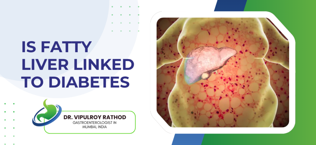
Yes, fatty liver and diabetes are directly linked through insulin resistance, the same metabolic dysfunction that drives fat accumulation in the liver also impairs glucose regulation, both conditions develop together and each makes the other worse, Type 2 diabetics have significantly higher rates of MASLD than the general population and people with fatty liver carry elevated diabetes risk even when their blood sugar currently looks fine on a report. According to Dr. Vipulroy Rathod, Gastroenterologist in Mumbai, “Fatty liver and diabetes are two expressions of the same underlying metabolic problem and treating one without addressing the other is why so many patients cycle through management plans that control numbers on paper without actually changing what’s happening in the liver.” https://www.youtube.com/shorts/i2roa-d-Ojs How Are Fatty Liver and Diabetes Connected? Same root cause, two organs expressing it differently, and most patients don’t find out the connection exists until both have been managed separately for years without either improving properly. Resistance: Liver cells stop responding to insulin, accumulate fat, the pancreas compensates by producing more which drives further fat storage, and that loop runs quietly until blood sugar can no longer be maintained regardless of how much insulin gets produced. Bidirectional: Fatty liver drives insulin resistance which drives diabetes, but existing diabetes accelerates liver fat accumulation and fibrosis progression simultaneously, so both get worse together unless both get properly managed together and most treatment plans don’t do that. MASH: Type 2 diabetics with fatty liver are far more likely to progress to metabolic steatohepatitis than fatty liver patients without diabetes, and most of them have no idea their diabetes is actively changing their liver disease trajectory while they’re managing blood sugar as a standalone problem. Fat: Central obesity deposits fat around internal organs including the liver, drives the inflammatory signals that damage liver tissue and impair beta cell function at the same time, and dietary advice given without structured metabolic intervention rarely reverses this in practice regardless of how motivated the patient is at the start. Managing fatty liver without looking at the metabolic picture underneath is treating a symptom not a disease. Specialist in fatty liver assesses the full metabolic context rather than just noting what the scan shows and stopping there. How Should Both Conditions Be Managed Together? Treating this as one metabolic problem rather than two separate referrals is what changes what management actually achieves for these patients. Weight: 5 to 10% body weight reduction improves liver fat, reduces fibrosis risk, and improves insulin sensitivity at the same time, and this is why structured weight loss works where generic lifestyle advice given at the end of a consultation consistently doesn’t. Medication: GLP-1 receptor agonists improve blood sugar, drive meaningful weight loss, and reduce liver fat through one mechanism rather than requiring separate drug regimens for two conditions that most patients can’t realistically maintain long term anyway. Monitoring: Both conditions together means fibrosis staging through fibroscan or FIB-4 blood markers, not just a liver ultrasound that tells the patient fat is present without telling them where on the fibrosis spectrum they actually sit or how urgently that matters. Alcohol: Even moderate drinking in a patient with both fatty liver and diabetes accelerates liver damage significantly beyond what either condition does alone, and most patients underestimate this because nobody tells them directly that their threshold is much lower than someone without their metabolic profile. Both conditions share the same root and addressing that root changes the trajectory of both. Read more on MASLD to understand the full picture of metabolic fatty liver disease and what proper management actually looks like. Why Choose Dr. Vipulroy Rathod Dr. Vipulroy Rathod has been managing fatty liver and its metabolic connections including diabetes and insulin resistance for over 30 years at Fortis Hospital Mulund, staging liver disease through fibroscan and blood markers rather than stopping at the ultrasound, building management plans that address the metabolic drivers rather than monitoring the liver while the underlying problem continues, trained physicians from 35 countries in this. Patients arrive having been told separately that they have fatty liver and that they have diabetes and most leave understanding for the first time that these are two expressions of one problem with one plan that actually addresses both. Start Your Treatment Journey Today Book Appointment Call now Frequently Asked Questions Does diabetes cause fatty liver? Yes, insulin resistance in Type 2 diabetes drives fat accumulation in the liver and significantly increases MASLD risk and progression rate. Can treating fatty liver improve diabetes control? Yes, reducing liver fat through weight loss and medication improves insulin sensitivity and often leads to better blood sugar control in Type 2 diabetics. How common is fatty liver in diabetic patients? Around 50 to 75% of Type 2 diabetic patients have some degree of fatty liver making it one of the most common complications of diabetes. Should diabetic patients get liver fibrosis testing? Yes, diabetic patients with fatty liver should have fibrosis staging through fibroscan or FIB-4 blood markers not just ultrasound given their higher progression risk. Reference links- Fatty Liver and Diabetes Connection — American Association for the Study of Liver Diseases MASLD and Metabolic Disease Guidelines — European Association for the Study of the Liver

Is Fatty Liver Linked to Diabetes Read More »

What Is EUS Celiac Plexus Neurolysis

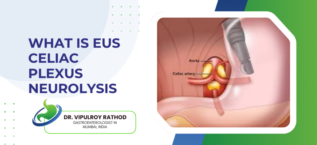
EUS celiac plexus neurolysis (CPN) is a minimally invasive procedure where alcohol is injected into the celiac plexus nerve cluster under endoscopic ultrasound guidance to permanently block pain signals from the pancreas, used primarily for pancreatic cancer pain that medication isn’t controlling, it works because it disrupts the nerve pathway at source rather than trying to suppress signals that keep firing regardless of what’s prescribed. According to Dr. Vipulroy Rathod, an experienced Gastroenterologist in Mumbai, “Celiac plexus neurolysis through EUS is one of the most impactful interventions available for pancreatic cancer pain because it targets the nerve pathway itself rather than just masking the pain with medication that stops working as disease progresses.” What Do Patients Actually Experience Before and After? Understanding the procedure honestly changes the decision-making for patients and families in ways that vague reassurance never does. Procedure: Sedation, scope into stomach, EUS finds the celiac plexus in real time, injection takes a few minutes once position is confirmed, total time 30 to 45 minutes, home same day and most people are surprised by how straightforward it actually was compared to what they imagined. Relief: Pain reduction within 24 to 72 hours in most cases, not all patients get complete relief but most get significant improvement, and the reduction in opioid dose that follows often resolves the nausea and constipation that heavy pain medication was causing on top of everything else. Risks: Temporary pain flare or diarrhoea in first 48 hours, hypotension occasionally, serious complications like paralysis are rare but real and this procedure should only be done by someone who’s performed enough of them to handle whatever comes up without sending the patient to a different team. Repeat: One session handles most patients for months, pain eventually returns as disease progresses, repeat neurolysis or transition to other palliative strategies decided based on how long the first response held and where the overall clinical picture sits at that point. This procedure doesn’t treat the cancer but it gives patients their life back during a period when that matters more than most people outside the situation understand. Read more on pancreatitis to see how EUS-guided intervention handles pancreatic conditions beyond just cancer pain relief. Why Does MASLD Diagnosis Actually Matter? Finding fat on a liver scan is where the investigation starts, not where it ends, and most patients have no idea what comes next. Stage: Simple steatosis carries low progression risk while MASH carries real fibrosis risk, and without staging through fibroscan, blood markers, or biopsy the patient has no idea which end of the spectrum they’re on or how urgently they need to act on it. Metabolic: MASLD means the underlying drivers, obesity, diabetes, insulin resistance, dyslipidaemia, need active management not just a lifestyle leaflet handed over at the end of a consultation nobody follows up on because nobody made it a clinical priority. Progression: Around 20% of MASLD patients with steatohepatitis progress to significant fibrosis over time and a proportion reach cirrhosis and liver cancer risk, so the patient told they have fatty liver and sent home without staging is in a completely different position from the patient properly managed from the start. Cardiovascular: MASLD carries significant cardiovascular risk independent of liver disease because the metabolic syndrome driving liver fat accumulation is the same syndrome driving heart disease, and patients with MASLD die of cardiovascular events more often than liver complications in early stage disease. Fatty liver report without staging and metabolic assessment is an incomplete workup that leaves the patient guessing. Read more on diagnosis to understand how the right investigation changes what gets found and what gets done about it. Why Choose Dr. Vipulroy Rathod Dr. Vipulroy Rathod has been performing EUS-guided celiac plexus neurolysis for over 30 years at Fortis Hospital Mulund, with enough case volume to know where the procedure works well, where it doesn’t, and what the realistic expectations are for each patient rather than offering blanket reassurance, trained physicians from 35 countries in exactly this. Patients arrive on heavy opioid loads in significant pain and most leave with a picture that looks completely different within days, not because the cancer changed but because the nerve driving the pain was finally addressed directly. Start Your Treatment Journey Today Book Appointment Call now Frequently Asked Questions Who is a candidate for EUS celiac plexus neurolysis? Patients with unresectable pancreatic cancer causing severe pain not controlled by oral analgesics are the primary candidates for this procedure. How long does pain relief last after EUS celiac plexus neurolysis? Pain relief typically lasts weeks to months and can be repeated if pain returns as disease progresses over time. Is EUS celiac plexus neurolysis safe? Yes, generally safe when performed by an experienced endoscopist with serious complications rare though temporary diarrhoea and hypotension can occur. What is the difference between celiac plexus neurolysis and celiac plexus block? Neurolysis uses alcohol for permanent nerve destruction while block uses steroids or local anaesthetic for temporary relief lasting weeks rather than months. Reference links- EUS Celiac Plexus Neurolysis Guidelines — American Society for Gastrointestinal Endoscopy Pancreatic Cancer Pain Management — World Gastroenterology Organisation

What Is EUS Celiac Plexus Neurolysis Read More »

What Is MASLD vs NAFLD

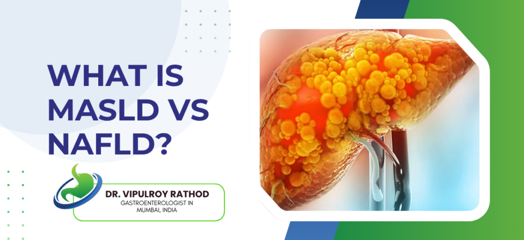
MASLD (Metabolic Dysfunction-Associated Steatotic Liver Disease) is the new term replacing NAFLD (Nonalcoholic Fatty Liver Disease), shifting focus from “not alcohol” to identifying the actual metabolic causes driving the disease. Both refer to fatty liver but MASLD requires at least one of five cardiometabolic risk factors including diabetes, high blood pressure, obesity, raised triglycerides, or low HDL, making diagnosis more precise and less stigmatising for patients. According to Dr. Vipulroy Rathod, an experienced, Gastroenterologist in Mumbai, “The name change from NAFLD to MASLD matters clinically because it shifts focus to the actual metabolic causes driving liver fat accumulation rather than just excluding alcohol, and that changes how patients understand and engage with their own risk.” What Is the Difference Between MASLD and NAFLD? Same liver condition, different name, and the reason behind the change matters more than most patients realise when they see both terms on different reports. Name: NAFLD defined the condition by absence of alcohol while MASLD defines it by presence of metabolic dysfunction, shifting clinical focus toward obesity, diabetes, and insulin resistance as primary targets rather than just watching the liver without addressing what’s actually driving the fat. Criteria: MASLD requires at least one cardiometabolic risk factor alongside liver steatosis while NAFLD simply required no significant alcohol without specifying what was actually causing the problem, which meant patients got a label without a proper metabolic workup attached to it. Spectrum: Both terms cover the same disease range from simple steatosis through steatohepatitis to fibrosis, cirrhosis, and liver cancer, and a scan showing fat is not the end of the investigation because where on that spectrum the patient sits determines everything that happens next. Adoption: MASLD is accepted terminology across major liver societies including EASL and AASLD from 2023, so patients seeing NAFLD on older reports and MASLD on newer ones are looking at the same condition under different naming conventions that changed mid-decade. Understanding what the name on your report actually means changes what investigation comes next. Specialist in fatty liver stages the disease properly rather than stopping at the scan finding and sending patients home without a clear picture of where they actually sit. Why Does MASLD Diagnosis Actually Matter? Finding fat on a liver scan is where the investigation starts, not where it ends, and most patients have no idea what comes next. Stage: Simple steatosis carries low progression risk while MASH carries real fibrosis risk, and without staging through fibroscan, blood markers, or biopsy the patient has no idea which end of the spectrum they’re on or how urgently they need to act on it. Metabolic: MASLD means the underlying drivers, obesity, diabetes, insulin resistance, dyslipidaemia, need active management not just a lifestyle leaflet handed over at the end of a consultation nobody follows up on because nobody made it a clinical priority. Progression: Around 20% of MASLD patients with steatohepatitis progress to significant fibrosis over time and a proportion reach cirrhosis and liver cancer risk, so the patient told they have fatty liver and sent home without staging is in a completely different position from the patient properly managed from the start. Cardiovascular: MASLD carries significant cardiovascular risk independent of liver disease because the metabolic syndrome driving liver fat accumulation is the same syndrome driving heart disease, and patients with MASLD die of cardiovascular events more often than liver complications in early stage disease. Fatty liver report without staging and metabolic assessment is an incomplete workup that leaves the patient guessing. Read more on diagnosis to understand how the right investigation changes what gets found and what gets done about it. Why Choose Dr. Vipulroy Rathod Dr. Vipulroy Rathod has been managing fatty liver disease and its metabolic drivers for over 30 years at Fortis Hospital Mulund, staging MASLD properly through fibroscan, blood markers, and EUS where indicated rather than stopping at the ultrasound finding and sending patients home with generic advice that changes nothing in practice, trained physicians from 35 countries in this approach. Patients arrive with a fatty liver report and no idea where they sit on the disease spectrum and most leave with a clear stage, a metabolic plan, and an actual follow-up schedule rather than a vague suggestion to eat better. Start Your Treatment Journey Today Book Appointment Call now Frequently Asked Questions Is MASLD the same as NAFLD? Yes, MASLD replaced NAFLD in 2023 as the globally accepted term for fatty liver disease driven by metabolic dysfunction. Can MASLD progress to liver cancer? Yes, MASLD can progress through steatohepatitis and fibrosis to cirrhosis and eventually hepatocellular carcinoma in a proportion of patients. What causes MASLD? Obesity, Type 2 diabetes, insulin resistance, high triglycerides, and high blood pressure are the primary metabolic drivers of MASLD. How is MASLD diagnosed and staged? Ultrasound or fibroscan identifies liver fat and fibrosis, blood markers like FIB-4 help stage disease, and biopsy confirms grade in uncertain cases. Reference links- MASLD Nomenclature and Definition — European Association for the Study of the Liver Fatty Liver Disease Guidelines — American Association for the Study of Liver Diseases

What Is MASLD vs NAFLD Read More »

How Is GI Bleeding Treated Without Surgery

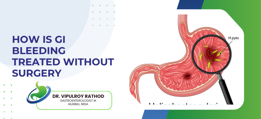
GI bleeding is often treated without surgery using endoscopic techniques such as clipping, heat cauterisation, or medication injection to seal bleeding vessels. For upper GI bleeds, intravenous proton pump inhibitors (PPIs) help reduce acid and support healing, while lower GI bleeding may be managed with medication or interventional radiology when needed. According to Dr. Vipulroy Rathod, Gastroenterologist in Mumbai, “GI bleeding is one of those emergencies where endoscopic techniques have advanced to the point where most cases that would have needed surgery fifteen years ago are now handled entirely inside the scope without a single incision.” What Endoscopic Treatments Stop GI Bleeding Without Surgery? Different sources need different tools and matching the right one to the right bleeding point matters more than just getting a scope in fast. Injection Therapy: Adrenaline injected into and around a bleeding ulcer causes vasoconstriction and tamponade that stops active bleeding quickly, almost always combined with a second modality like clipping because injection alone carries higher rebleeding rates than combination treatment. Endoscopic Clipping: Mechanical clips applied directly to a bleeding vessel close it permanently without heat or chemical, particularly effective for Dieulafoy lesions, visible vessels in ulcer bases, and Mallory-Weiss tears where precision matters more than thermal spread. Argon Plasma Coagulation: Non-contact thermal coagulation that treats vascular lesions, radiation-induced proctitis, gastric antral vascular ectasia, and diffuse mucosal bleeding across wide surface areas where clipping or injection simply isn’t practical. Band Ligation for Varices: Oesophageal varices from portal hypertension causing catastrophic upper GI bleeding controlled through band ligation in the acute setting and then through elective sessions to obliterate remaining varices and prevent recurrence. Right tool for right source changes outcomes and patients at experienced endoscopy centres reach surgery far less often than those where endoscopic technique is limited. Proper endoscopy handles the full range of GI bleeding without defaulting to surgical referral when the endoscopic solution exists. When Is Surgery Actually Needed for GI Bleeding? Minority of cases, but real situations exist and knowing when to stop attempting endoscopy matters as much as knowing how to do it. Haemodynamic Instability Despite Endoscopy: Massive ongoing haemorrhage where the patient can’t be stabilised despite resuscitation and endoscopic attempts, surgical exploration becomes the right call and delaying it for another scope session costs time the patient doesn’t have. Endoscopic Access Failure: Bleeding from jejunum or proximal ileum beyond scope reach, anatomical distortion from previous surgery preventing access to the bleeding point, these need surgical intervention because the scope physically cannot get where it needs to be. Aortoenteric Fistula: Bleeding from a vascular graft eroding into bowel is a surgical emergency regardless of endoscopic findings because the underlying vascular problem cannot be addressed through the scope no matter how experienced the endoscopist is. Recurrent Bleeding Despite Two Attempts: Two failed endoscopic attempts at the same bleeding source means the vessel is too large or lesion too complex for endoscopic haemostasis and surgical ligation gives more durable control than a third session with the same likely outcome. Most GI bleeding gets sorted endoscopically when the right person is doing it and the right tools are available. Read more on procedures to understand what’s possible without surgery across the full range of GI conditions. Why Choose Dr. Vipulroy Rathod Dr. Vipulroy Rathod has been managing acute and chronic GI bleeding through injection therapy, clipping, argon plasma coagulation, and variceal band ligation for over 30 years at Fortis Hospital Mulund, with a case volume and endoscopic range that means bleeding sources other endoscopists send to surgery get controlled here without an incision in most cases, trained physicians from 35 countries in exactly this. Patients arrive in active bleed referred for surgical opinion and most leave having had the bleeding controlled endoscopically the same day without ever meeting a surgeon. Start Your Treatment Journey Today Book Appointment Call now Frequently Asked Questions What is the most common cause of upper GI bleeding treated without surgery? Peptic ulcer bleeding is the most common cause of upper GI bleeding and is successfully managed endoscopically in the majority of cases. How quickly does endoscopic treatment stop GI bleeding? Active GI bleeding is typically controlled within the same endoscopic session, usually within 30 to 60 minutes of the procedure starting. Can variceal bleeding be permanently treated without surgery? Yes, repeated endoscopic band ligation sessions obliterate oesophageal varices and prevent recurrent bleeding without surgical intervention in most patients. What happens if endoscopy fails to stop GI bleeding? If endoscopy fails after two attempts, interventional radiology embolisation or surgical exploration becomes the next step depending on source and patient stability. Reference links- Endoscopic Management of GI Bleeding — American Society for Gastrointestinal Endoscopy GI Bleeding Treatment Guidelines — World Gastroenterology Organisation

How Is GI Bleeding Treated Without Surgery Read More »

Can Chronic Pancreatitis Be Treated Without Surgery

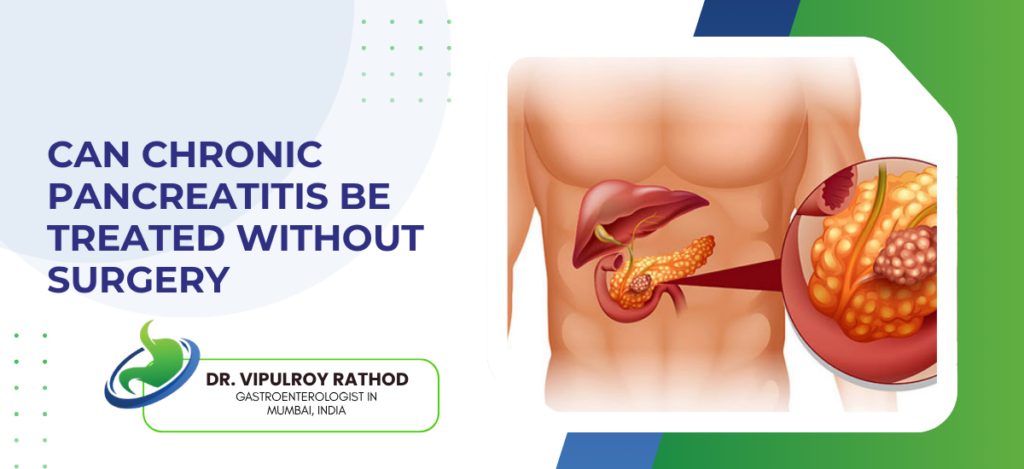
Yes, chronic pancreatitis can often be managed without surgery through lifestyle changes, medications, and endoscopic procedures. The main goals are to control pain, improve digestion, and manage diabetes when present. Common non-surgical treatments include pain relief medication, pancreatic enzyme supplements, strict avoidance of alcohol and tobacco, and a low-fat diet. According to Dr. Vipulroy Rathod, Gastroenterologist in Mumbai, “Chronic pancreatitis is a condition where the right non-surgical management early on prevents the kind of disease progression that eventually forces surgical intervention, and most patients who end up needing surgery got there because endoscopic and medical options were not used properly or early enough.” https://www.youtube.com/shorts/ZXWA_aRaVQ4 What Non-Surgical Treatments Actually Work Here? Several. Sequence matters more than most people realise and getting it wrong early creates the surgical case that didn’t need to exist. Pain at Root Cause: Ductal hypertension drives most chronic pancreatitis pain and decompressing it endoscopically changes the trajectory completely for patients where obstruction is the driver, not just neural inflammation that medication alone won’t fix anyway. Enzyme Replacement: As pancreatitis destroys enzyme-producing tissue exocrine insufficiency develops and oral replacement corrects malabsorption, improves nutrition, and reduces the postprandial pain most patients are blaming on the pancreatitis itself when it’s actually the deficiency underneath. ERCP for Ductal Stones: Stones and strictures removed or dilated endoscopically, most patients discharged within 48 hours, no incision, and a proportion of them report more pain relief from this single intervention than from months of medication that was addressing the symptom not the cause. EUS-Guided Pseudocyst Drainage: Fluid collections causing nausea and gastric outlet symptoms drained transmurally without surgery and patient avoids surgical recovery entirely for what sounds like a complex problem but isn’t when done by someone who does it regularly. Non-surgical management works when matched properly to the presentation rather than defaulting to surgical referral because it’s the easier path. A proper treatment plan sequences these correctly without skipping steps. When Does Surgery Actually Become Necessary? Minority of cases. But real situations exist and recognising them matters as much as knowing when to avoid surgery. Endoscopy Reaches Its Limits: Stones too impacted for ERCP even with lithotripsy, strictures too complex for stenting, pseudocysts in positions not accessible transmurally  these move to surgical discussion but genuinely represent a small fraction of cases at centres with real endoscopic range. Malignancy Can’t Be Excluded: When a mass develops in the context of chronic pancreatitis that EUS biopsy can’t call benign definitively, surgery becomes diagnostic and potentially curative and delaying it to repeat non-surgical attempts costs progression time nobody recovers. Structural Complications: Splenic artery pseudoaneurysm bleeding, splenic vein thrombosis causing portal hypertension, pancreatic head fibrosis not responding to stenting endoscopy doesn’t fix structural problems regardless of who’s doing it. Pain That Didn’t Respond to a Real Attempt: Patients who’ve genuinely had proper endoscopic intervention, enzyme replacement, dietary modification, and optimised pain management without relief are surgical candidates, but that sequence needs to have actually happened properly first. Most chronic pancreatitis doesn’t reach surgery when the right specialist manages it from the start, and regular surveillance for malignant change is part of the same picture not a separate conversation. Why Choose Dr. Vipulroy Rathod Dr. Vipulroy Rathod has been managing chronic pancreatitis through ERCP, EUS-guided drainage, and endoscopic ductal intervention for over 30 years at Fortis Hospital Mulund, keeping patients out of the operating theatre through properly sequenced non-surgical treatment that most gastroenterologists refer to surgeons too early, trained physicians from 35 countries in exactly this. Patients arrive having been told surgery is the only option and most leave with a plan that addresses what’s actually driving the symptoms rather than managing around them. Start Your Treatment Journey Today Book Appointment Call now Frequently Asked Questions Can chronic pancreatitis pain be controlled without surgery? Yes, endoscopic ductal decompression, enzyme replacement, and targeted pain management control chronic pancreatitis pain without surgery in most patients. How long does ERCP treatment for chronic pancreatitis take? ERCP for pancreatic duct stones or strictures takes 45 to 90 minutes under sedation with most patients discharged the following day. Can exercise reduce colorectal cancer risk in genetically predisposed patients? No, but chronic pancreatitis increases lifetime pancreatic cancer risk and requires regular EUS surveillance to detect malignant change early.   Can diet changes help chronic pancreatitis without surgery? Yes, low-fat diet, alcohol cessation, small frequent meals, and pancreatic enzyme supplementation significantly reduce symptom burden without surgical intervention. Reference links- Chronic Pancreatitis Management Guidelines — American College of Gastroenterology Non-Surgical Pancreatitis Treatment — World Gastroenterology Organisation

Can Chronic Pancreatitis Be Treated Without Surgery Read More »

What Is Digestive Cancer Treatment Cost in Mumbai 2026

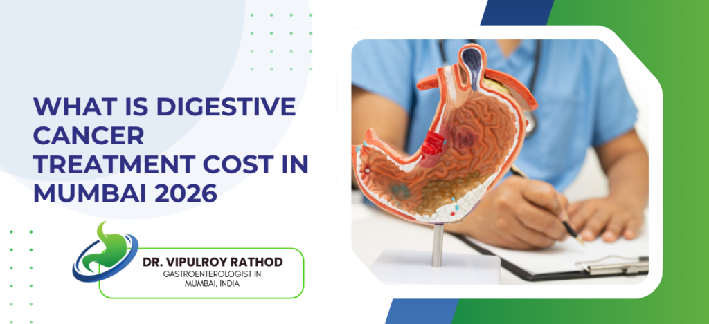
Digestive cancer treatment cost in Mumbai in 2026 starts at Rs 80,000 for early endoscopic Digestive cancer treatment costs in Mumbai in 2026 generally range from ₹2,25,000 to over ₹10,00,000, depending largely on the cancer stage, treatment approach such as surgery, chemotherapy, or radiation, and the hospital selected. Surgical treatment commonly costs around ₹3–4.5 lakh, while chemotherapy may range from approximately ₹2–8 lakh. According to Dr. Vipulroy Rathod, Gastroenterologist in Mumbai, “The most expensive digestive cancer treatment is always late-stage disease, finding it early doesn’t just improve survival, it reduces treatment complexity and cost significantly and that is a clinical and financial reality most patients don’t consider until they’re already in the middle of it.” What Actually Drives the Cost Up? Stage at diagnosis matters more than hospital name, surgeon reputation, or which treatment protocol gets chosen. Stage: Same colorectal cancer at Stage 1 costs a fraction of what Stage 3 demands with surgery plus months of chemotherapy stacked on top, and most patients only discover this gap exists after they’ve already arrived at the wrong stage. Approach: Endoscopic removal costs far less than surgery, shorter stay, no ICU, quicker recovery, but this option disappears once cancer has grown past the stage where the scope can handle it without a knife getting involved. Chemotherapy: Rs 60,000 to Rs 1,50,000 per cycle, up to 12 cycles for Stage 3 colorectal cancer, and patients are often blindsided by how the scan costs, supportive medications, and outpatient visits quietly add to the main treatment bill over months. Hospital: High-volume centres charge more upfront but the real financial risk with cheaper low-volume options is complications and repeat procedures that nobody quotes in the initial bill. Early detection keeps the cost in a range where treatment is actually curative, specialist in cancer catches it early enough for those options to still exist rather than managing advanced disease after the window has already closed. What Does Each Treatment Actually Cost in Mumbai 2026? Ballpark figures only, real cost varies with case complexity, hospital, and how cleanly the procedure goes. Endoscopic: Rs 80,000 to Rs 2,50,000 for early mucosal stomach or oesophageal cancers, day procedure, discharged same day or next morning, and the only patients who reach this option are the ones who got investigated before the cancer outgrew it. Colorectal: Laparoscopic colectomy Rs 3,50,000 to Rs 6,00,000, chemotherapy for Stage 3 at Rs 60,000 to Rs 1,50,000 per cycle over 6 to 12 cycles, total episode cost crossing Rs 15,00,000 before supportive care gets factored in. Pancreatic: Whipple procedure Rs 6,00,000 to Rs 15,00,000, ICU standard, adjuvant chemotherapy adds more, and outcomes here depend heavily on which centre does it and how many of these they actually do per year. Palliative: Bile duct stenting Rs 80,000 to Rs 1,50,000, EUS-guided drainage Rs 1,00,000 to Rs 2,00,000, these manage advanced disease without surgery at significantly lower hospitalisation cost when cure is no longer realistic. Treatment picture changes completely when disease is caught early and the endoscopic option is still available. Read more on procedures to understand what endoscopic treatment delivers compared to surgical alternatives. Why Choose Dr. Vipulroy Rathod Dr. Vipulroy Rathod has been performing endoscopic resection, EUS-guided intervention, and therapeutic endoscopy for over 30 years at Fortis Hospital Mulund, consistently giving patients access to curative endoscopic treatment that avoids surgery entirely when disease is caught at the right stage, trained physicians from 35 countries in doing exactly this rather than defaulting to surgical referral when the endoscopic option still exists. Patients who come in early spend less, recover faster, and leave wit Start Your Treatment Journey Today Book Appointment Call now Frequently Asked Questions How much does colorectal cancer treatment cost in Mumbai? Laparoscopic colorectal surgery costs Rs 3,50,000 to Rs 6,00,000 with chemotherapy adding Rs 60,000 to Rs 1,50,000 per cycle for advanced stages. Is endoscopic cancer treatment cheaper than surgery in Mumbai? Yes, endoscopic resection for early GI cancers costs Rs 80,000 to Rs 2,50,000 compared to Rs 3,00,000 to Rs 15,00,000 for surgical approaches. Can exercise reduce colorectal cancer risk in genetically predisposed patients? Does health insurance cover digestive cancer treatment in Mumbai?Most health insurance policies cover surgery and chemotherapy but coverage for endoscopic procedures varies significantly by policy and insurer. What is the cheapest way to treat digestive cancer in Mumbai? Early detection allowing endoscopic resection is most cost-effective, avoiding surgery, ICU admission, and prolonged chemotherapy entirely. Reference links- GI Cancer Treatment Guidelines India — Indian Council of Medical Research Digestive Cancer Management Costs — World Gastroenterology Organisation

What Is Digestive Cancer Treatment Cost in Mumbai 2026 Read More »

Can Healthy Habits Reduce Genetic Risk of Digestive Cancer

Yes, healthy habits can significantly help reduce the risk of digestive cancers, even in people with a genetic predisposition. Maintaining a healthy weight, exercising regularly, eating fibre-rich foods, and avoiding smoking and alcohol can lower risk by reducing inflammation and limiting long-term cellular damage. According to Dr. Vipulroy Rathod, an experienced, Gastroenterologist in Mumbai, “Genetic risk is real but it is not a fixed sentence, patients with strong family history who come in for surveillance regularly and make the right lifestyle changes give themselves a meaningfully different clinical picture than those who assume the outcome is already decided.” How Do Healthy Habits Reduce Genetic Risk of Digestive Cancer? Genes set the starting point. What happens next is not fixed. Diet Changes Gene Expression: High fibre diet reduces colorectal cancer risk even in Lynch syndrome carriers by changing gut microbiome composition and reducing bile acid concentration in the colon, red and processed meat pushes genetically susceptible cells toward malignancy faster by producing carcinogenic compounds during digestion. Exercise:Physical activity cuts colorectal cancer risk by 20 to 30% in population studies. Not 2 to 3%. Twenty to thirty. For someone already at genetic risk that is not a marginal number, it shifts the entire clinical picture and most patients are never told this directly. Tobacco and Alcohol Multiply Risk, Not Add: Both are independent carcinogens and in someone with BRCA2 or Lynch syndrome they don’t just add to existing risk, they multiply it, quitting both removes the environmental accelerant from a system already predisposed to malignant change. MetabolicRisk: BMI above 30 in a Lynch syndrome carrier that combination warrants aggressive surveillance, not just lifestyle advice at the end of a routine consultation that the patient forgets by the time they reach the car park. Lifestyle changes reduce risk. Don’t eliminate it. Specialist in GI cancer treatment combines both rather than treating them as separate conversations that happen in different rooms. What Surveillance Should High-Risk Patients Actually Get? Healthy habits and surveillance work together. One without the other leaves gaps that matter clinically. Lynch Syndrome, Not Every 10 Years: Standard population colonoscopy interval is 10 years, Lynch syndrome carriers need it every 1 to 2 years from age 20 to 25, polyps removed at that frequency never get the chance to become the cancer their genetics predicted. BRCA2 mutation significantly elevates pancreatic cancer risk. Annual EUS surveillance for BRCA2 carriers finds pancreatic lesions at resectable stage. Most of these patients have zero symptoms when the lesion is found. That’s the whole point of surveillance and it only works if someone actually orders it. Family History Without Known Mutation: One first-degree relative with colorectal cancer means colonoscopy starting 10 years before that relative’s diagnosis age, two relatives or one diagnosed under 50 means surveillance as aggressive as Lynch syndrome regardless of what the genetic test shows. Upper GI surveillance for gastric cancer risk gets skipped constantly in India. Patients with family history of stomach cancer or H. pylori positive status in high-incidence families need regular upper endoscopy, finding early gastric cancer at mucosal stage means endoscopic resection without surgery. Genetics is the starting point. Surveillance changes the ending. Read more on FASGE fellowship to understand the expertise behind this surveillance approach. Why Choose Dr. Vipulroy Rathod Dr. Vipulroy Rathod has been evaluating pancreatic cysts through EUS since 1998, with over 30 years of experience differentiating benign cysts from those with malignant potential using fine needle aspiration, cyst fluid analysis, and EUS morphology that CT cannot replicate. Trained physicians from 35 countries in exactly this pancreatic cyst assessment at Fortis Hospital Mulund. Patients arrive with a cyst report and no idea what it means. Most leave knowing exactly what type of cyst it is, whether it needs treatment, and what monitoring looks like if it doesn’t. Start Your Treatment Journey Today Book Appointment Call now Frequently Asked Questions Does diet alone prevent genetic digestive cancer risk? No, diet reduces risk significantly but must be combined with active surveillance through colonoscopy and EUS for high-risk patients. At what age should Lynch syndrome carriers start colonoscopy? Lynch syndrome carriers should start colonoscopy surveillance at age 20 to 25 or 10 years before the earliest family diagnosis. Can exercise reduce colorectal cancer risk in genetically predisposed patients? Yes, regular physical activity reduces colorectal cancer risk by 20 to 30% even in patients with genetic predisposition. Do BRCA2 mutation carriers need pancreatic cancer surveillance? Yes, annual EUS surveillance is recommended for BRCA2 carriers given their significantly elevated lifetime risk of pancreatic cancer. Reference links- Genetic Risk and GI Cancer Prevention — American College of Gastroenterology Hereditary GI Cancer Surveillance Guidelines — World Gastroenterology Organisation

Can Healthy Habits Reduce Genetic Risk of Digestive Cancer Read More »

Pancreatic Cancer vs Pancreatic Cyst: What Is the Difference

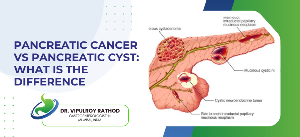
Pancreatic cysts are fluid-filled sacs in or on the pancreas, most are benign and found incidentally on imaging, but some carry malignant potential and need monitoring or treatment. Pancreatic cancer is solid malignant tumour growth in pancreatic tissue, aggressive, and life-threatening when found late. According to Dr. Vipulroy Rathod, an experienced, Gastroenterologist in Mumbai, “A pancreatic cyst report from a scan does not tell you whether you need to watch it, treat it, or remove it  that answer comes from EUS and the specialist reading it, not from the CT report alone.” How Are Pancreatic Cancer and Pancreatic Cysts Different? Same organ, completely different pathology. And the consequences of treating one like the other go in both directions. What They Are: Pancreatic cancer is solid tumour, malignant from the start, grows and invades surrounding tissue, pancreatic cyst is fluid-filled space, most are benign pseudocysts from prior pancreatitis or serous cystadenomas that carry virtually no cancer risk at all. How They’re Found: Cancer usually found because patient has symptoms, weight loss, jaundice, abdominal pain, cysts found incidentally on CT or MRI done for something completely unrelated, patient had no symptoms, no idea the cyst was there. Which Cysts Actually Worry Specialists: Serous cystadenomas essentially benign, pseudocysts benign in context of pancreatitis, mucinous cystic neoplasms and IPMNs carry real malignant potential especially when cyst is large, has solid components, or shows worrying ductal features on EUS that plain CT simply doesn’t show. Symptoms: Pancreatic cancer causes progressive pain, weight loss, jaundice, new onset diabetes, most pancreatic cysts cause no symptoms at all and produce no clinical signs until they’re either very large or already transforming, which is exactly why active surveillance matters for the right cyst types. Not all cysts need treatment. Not all cysts are safe to ignore. Specialist in pancreatic cancer treatment knows exactly which category a cyst falls into and what the next step actually is When Does a Pancreatic Cyst Become a Concern? Most patients with incidental cysts are told to repeat the scan in six months. That’s not always the right answer. Size and Growth Rate: Cysts growing more than 5mm per year or already above 3cm at first detection need proper EUS evaluation not just a repeat CT, growth rate matters as much as absolute size and CT alone doesn’t give you the detail needed to assess wall changes. Solid Components Inside the Cyst: Mural nodule inside a cyst changes the risk profile significantly, that finding on EUS moves the clinical decision from surveillance to intervention discussion immediately regardless of cyst size. Ductal Involvement: Main pancreatic duct dilation alongside a cyst is a worrying combination, specifically in IPMN cases, and EUS with fine needle aspiration of cyst fluid for CEA and amylase levels gives information no external scan provides. Patient Risk Profile: Family history of pancreatic cancer, BRCA2 mutation carrier, chronic pancreatitis history alongside a cyst of uncertain type, that combination changes surveillance frequency and investigation intensity compared to a low-risk patient with a simple serous cyst. Finding a cyst on a scan is not reassurance. It’s the beginning of an investigation that needs the right specialist. Read more on pancreatic cyst treatment to understand what proper evaluation and management actually involves. Why Choose Dr. Vipulroy Rathod Dr. Vipulroy Rathod has been evaluating pancreatic cysts through EUS since 1998, with over 30 years of experience differentiating benign cysts from those with malignant potential using fine needle aspiration, cyst fluid analysis, and EUS morphology that CT cannot replicate. Trained physicians from 35 countries in exactly this pancreatic cyst assessment at Fortis Hospital Mulund. Patients arrive with a cyst report and no idea what it means. Most leave knowing exactly what type of cyst it is, whether it needs treatment, and what monitoring looks like if it doesn’t. Start Your Treatment Journey Today Book Appointment Call now Frequently Asked Questions Are all pancreatic cysts dangerous? No. Most pancreatic cysts are benign but certain types like IPMNs and mucinous cysts carry malignant potential and need proper surveillance. Can a pancreatic cyst turn into cancer? Yes, mucinous cystic neoplasms and main duct IPMNs have significant malignant potential and require regular EUS monitoring or surgical removal. How is a pancreatic cyst different from pancreatic cancer on imaging? Cysts appear as fluid-filled spaces on CT while cancer appears as a solid mass, but small cancers and complex cysts need EUS for accurate differentiation.   What test best evaluates a pancreatic cyst? EUS with fine needle aspiration and cyst fluid analysis for CEA and amylase levels gives the most accurate characterisation of pancreatic cysts. Reference links- Pancreatic Cyst Evaluation Guidelines — American College of Gastroenterology Pancreatic Cysts and Malignant Potential — World Gastroenterology Organisation

Pancreatic Cancer vs Pancreatic Cyst: What Is the Difference Read More »

Scroll to Top
Call Now Button