

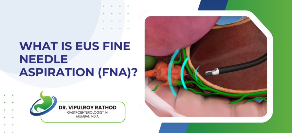
EUS Fine Needle Aspiration (EUS-FNA) is a minimally invasive biopsy procedure that combines endoscopic ultrasound with a thin needle to sample tissue from organs deep inside the abdomen and chest. It is the standard method for getting a definitive diagnosis from pancreatic lesions, lymph nodes, the bile duct wall, and submucosal masses in the GI tract. The whole thing happens through a scope passed down the mouth, no incisions, usually under sedation. For most pancreatic cancers, EUS-FNA is what confirms the diagnosis before any treatment decision gets made.
According to Dr. Vipulroy Rathod, Gastroenterologist in Mumbai, “EUS-FNA changed how we diagnose pancreatic disease. Before it existed, patients went straight to surgery on the basis of a CT scan. Now we sample the lesion first. The biopsy answers the question. Sometimes it isn’t cancer at all, and we’ve saved someone from a major operation they never needed.”
What EUS-FNA is used for?
The procedure is built around one capability, getting tissue from places that are hard to reach any other way. The pancreas sits behind the stomach, deep in the abdomen, and is impossible to biopsy through the skin in most cases without risk.
- Pancreatic masses. The classic indication. Any solid lesion in the pancreas needing histological confirmation before treatment planning. The needle goes through the stomach or duodenum wall directly into the pancreas, real-time ultrasound guidance the whole way.
- Lymph node sampling for staging in cancers of the oesophagus, lung, pancreas, and stomach. Knowing whether nearby nodes are involved changes the surgical plan, sometimes pulls a case from operable to inoperable.
- Cystic lesions of the pancreas. Fluid aspirated from a pancreatic cyst is sent for cytology, CEA, glucose, and now molecular markers. This is what tells the difference between a benign serous cyst and a mucinous one with cancer potential.
- Submucosal tumours of the stomach and oesophagus. These sit beneath the lining and can’t be reached with a regular biopsy through a standard endoscope. EUS-FNA goes through the wall and samples them directly.
The diagnostic yield depends on the operator, the needle, and the on-site cytology setup. Endoscopic ultrasound services that have a cytopathologist available during the procedure consistently produce better samples, because the operator gets immediate feedback on whether the tissue is adequate or another pass is needed.
How EUS-FNA is performed?
The procedure itself is quick, usually thirty to forty minutes from start to finish, and patients go home the same day in most cases.
A linear-array echoendoscope is passed through the mouth and positioned in the stomach or duodenum, depending on which organ is being sampled. The ultrasound at the tip of the scope locates the lesion, the operator measures it, and a fine needle, usually 22 or 25 gauge, is advanced through the scope and into the target under continuous ultrasound guidance.
- Multiple passes. Two to four passes through the lesion are standard, each pass collecting tissue into a syringe attached to the needle hub. The cytopathologist examines the smears in real time when ROSE (rapid on-site evaluation) is available.
- Most patients receive deep sedation rather than general anaesthesia. Recovery is short, an hour or two in the day-care unit, and the patient usually goes home by evening.
- Complications are rare. Bleeding, pancreatitis, and infection occur in well under one percent of cases in experienced hands. Tumour seeding along the needle tract is theoretically possible but extremely uncommon in pancreatic FNA.
- Results take three to five working days, sometimes faster if ROSE was used during the procedure to confirm adequacy.
The accuracy of EUS-FNA in pancreatic masses runs above ninety percent in expert centres, which is why it has become the standard step before any pancreatic surgery is scheduled. Read more on pancreatic cancer detection for how EUS-FNA fits into the broader workup of suspected pancreatic disease.
Why choose Dr. Vipulroy Rathod for EUS-FNA?
Dr. Vipulroy Rathod has been performing EUS-FNA at Fortis Hospital Mulund since the technique first became available in India in the late 1990s, which makes him one of the earliest endosonographers in the country to adopt it. Over thirty years of pancreatic biopsies, lymph node staging, cystic lesion characterisation, and the careful technique that makes each pass count.
What separates a good EUS-FNA from a poor one isn’t the equipment, it’s the operator. The choice of needle, the angle of approach, the number of passes, the handling of the specimen, all of these affect whether the cytopathologist gets enough tissue to give a definitive answer. Volume teaches that part of the work.
Book your consultation today with one of India’s most experienced endosonographers for EUS-FNA and pancreatic disease assessment.
Frequently Asked Questions
No, the procedure is done under sedation and patients don’t feel it. Mild throat soreness for a day afterwards is common, but most patients are eating normally by evening.
In experienced hands, diagnostic accuracy for pancreatic masses runs above ninety percent. False negatives can occur in small or fibrotic tumours, which is why a single negative biopsy doesn’t always rule out cancer if the imaging looks suspicious.
The procedure itself is usually thirty to forty minutes. Including sedation recovery, most patients are in the day-care unit for three to four hours before going home.
The theoretical risk of tumour seeding along the needle tract exists but is extremely rare in pancreatic FNA. For lesions that will be surgically resected, the needle tract is included in the resection specimen, eliminating any residual risk.
Reference links-
- EUS-Guided Tissue Acquisition Guidelines, American Society for Gastrointestinal Endoscopy — https://www.asge.org/home/practice-support/guidelines
- Endoscopic Ultrasound in Pancreatic Disease, World Gastroenterology Organisation — https://www.worldgastroenterology.org/guidelines
