

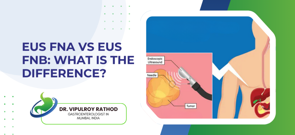
EUS Fine Needle Aspiration (FNA) and EUS Fine Needle Biopsy (FNB) are both endoscopic ultrasound-guided sampling procedures used to obtain tissue from lesions in the pancreas, lymph nodes, and submucosal masses. The difference is in what each needle collects, FNA aspirates loose cells suspended in fluid for cytology, while FNB captures a small intact tissue core that preserves the lesion’s architecture for histology. Both share the same setup, the same scope, the same patient experience, but the diagnostic yield differs in important ways depending on what’s being sampled.
According to Dr. Vipulroy Rathod, Gastroenterologist in Mumbai, “FNA was the original technique and still works well for most pancreatic masses. FNB came later. The newer needle pulls actual tissue, not just cells, which matters for lymphomas, autoimmune disease, neuroendocrine tumours, anything where the cytologist needs to see how the cells are arranged, not just what they look like individually.”
EUS-FNA vs EUS-FNB at a glance
| Factor | EUS-FNA | EUS-FNB |
| Sample type | Cells in fluid | Tissue core |
| Analysis | Cytology | Histology |
| Passes needed | Three to four | One to two |
| Best for | Solid pancreatic mass | Lymphoma, neuroendocrine |
| ROSE dependency | High | Low |
How the two techniques differ?
The needles themselves look similar from the outside, both pass through the same scope into the same target, both use the same ultrasound guidance. What changes is the tip design.
- FNA needle. A simple bevelled tip designed to aspirate cells and small fragments under suction. The sample comes out as a smear that’s spread on slides and stained for cytology. Multiple passes are usually needed, three or four, sometimes more if the cytopathologist isn’t on site to check adequacy in real time.
- FNB needles come in several designs, Franseen, fork-tip, reverse-bevel, all built to cut and retain a tissue core rather than just aspirate cells. One or two passes usually give enough tissue for histological analysis, which preserves the architecture of the lesion rather than just sampling individual cells.
- Diagnostic yield differs by target. For solid pancreatic adenocarcinoma, FNA and FNB perform similarly, both above ninety percent accuracy in expert hands. For lymphomas, gastrointestinal stromal tumours, autoimmune pancreatitis, and neuroendocrine tumours, FNB is meaningfully better because histology matters.
- Cost and availability also differ. FNA needles are cheaper and available in every endoscopy unit doing EUS. FNB needles cost more, aren’t stocked everywhere, and are sometimes used selectively rather than as the default.
What separates a successful biopsy from a non-diagnostic one is matching the technique to the lesion. Endoscopic ultrasound services that have both FNA and FNB available let the operator choose mid-procedure based on what the lesion looks like, rather than committing to one approach before the scope is even passed.
When each technique is preferred?
The choice isn’t always either-or. Many cases get FNA first, with FNB held in reserve if the initial passes are non-diagnostic.
- Solid pancreatic mass, likely adenocarcinoma. FNA is usually enough. Pancreatic cancer cells are recognisable on cytology, and the architecture matters less for diagnosis. ROSE confirms adequacy before the patient leaves the procedure room.
- For suspected lymphoma anywhere, FNB is preferred from the start. Lymphoma diagnosis needs flow cytometry, immunohistochemistry, and architectural assessment, none of which a cell aspirate can provide reliably. Going straight to FNB saves a repeat procedure.
- Subepithelial GI tumours. Stromal tumours, leiomyomas, schwannomas. These need histology and immunohistochemistry to differentiate. FNB is the default.
- Autoimmune pancreatitis is another situation where FNB matters. The diagnosis depends on seeing characteristic inflammatory patterns in the tissue, lymphoplasmacytic infiltrate, storiform fibrosis, obliterative phlebitis. None of these can be picked up reliably on FNA cytology.
So the decision often gets made in the procedure room itself, based on what the ultrasound is showing. Read more on pancreatic cancer detection to understand how the choice of biopsy technique fits into the broader workup of suspected pancreatic disease.
Why choose Dr. Vipulroy Rathod for EUS biopsy?
Dr. Vipulroy Rathod has been performing EUS-guided tissue sampling at Fortis Hospital Mulund since the technique first arrived in India, with experience covering both FNA and the newer FNB needles as they became available over the years. Over three decades of pancreatic masses, lymph node staging, cystic lesion analysis, and the case-by-case judgment that decides which needle gets used.
The needle choice matters more than patients usually realise. Wrong technique on the wrong lesion produces a non-diagnostic biopsy, which means a repeat procedure, more sedation, more delay. Right technique on the first attempt avoids that whole detour.
Book your consultation today with one of India’s most experienced endosonographers for EUS-guided biopsy and pancreatic disease assessment.
Frequently Asked Questions
For most solid pancreatic masses, accuracy is similar between the two. For lymphomas, neuroendocrine tumours, GI stromal tumours, and autoimmune pancreatitis, FNB is meaningfully better because the diagnosis needs histology rather than cytology alone.
No. The procedure feels the same from the patient’s side. Same sedation, same scope, same recovery time. The difference is in the needle tip and what comes out at the end.
Yes, often. Many operators start with FNA, check adequacy with ROSE if available, and switch to FNB if the initial passes don’t give enough material. Both needles fit through the same scope.
Cost, availability, and the fact that FNA still works well for the most common indication, solid pancreatic adenocarcinoma. In centres where both are available, FNB use has been steadily rising for lesions where histology matters.
Reference links-
- EUS-Guided Tissue Acquisition Guidelines, American Society for Gastrointestinal Endoscopy — https://www.asge.org/home/practice-support/guidelines
- Endoscopic Ultrasound in Pancreatic Disease, World Gastroenterology Organisation — https://www.worldgastroenterology.org/guideline
-
