

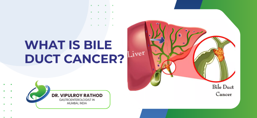
Bile duct cancer, also called cholangiocarcinoma, is a rare and aggressive malignancy of the cells lining the bile ducts. It can arise anywhere along the biliary tree, from inside the liver down to where the duct enters the duodenum. The classification follows location, intrahepatic, perihilar, distal. Most patients present late because the early disease is silent. Jaundice. Weight loss. Itching that won’t go away. By the time these appear, treatment options have already narrowed.
According to Dr. Vipulroy Rathod, Gastroenterologist in Mumbai, “Cholangiocarcinoma is hard to catch early. The ducts sit deep in the abdomen. Imaging shows duct dilatation. Often it doesn’t show the tumour itself. Patients arrive having been worked up for hepatitis or gallstones for weeks before anyone thinks of cancer. By then, resectability has often already gone.”
What are the types of cholangiocarcinoma and how do they present?
Classification follows location along the biliary tree. Location dictates symptoms, workup, and what treatment is possible.
- Intrahepatic cholangiocarcinoma. Inside the liver. Often a liver mass on imaging done for something else.
- Perihilar is the commonest type. Sits at the junction where the right and left hepatic ducts join, also called a Klatskin tumour. Presents as painless obstructive jaundice when the tumour blocks bile flow, and is often the hardest type to fully resect because of the proximity to major vascular structures and the need for extensive liver and bile duct resection together.
- Distal cholangiocarcinoma. Lower bile duct as it passes through the pancreas. Mimics pancreatic head cancer in presentation, gets worked up the same way, and the Whipple procedure is the operation when resectable.
- Risk factors matter but only in a minority. Primary sclerosing cholangitis. Choledochal cysts. Chronic biliary infection. Hepatitis B and C. Most cases occur in patients without any identifiable risk factor at all, which is part of what makes early detection so difficult and why surveillance is currently limited to high-risk groups rather than the general population.
The workup needs both imaging and tissue. Endoscopic ultrasound with fine-needle aspiration is often the only way to obtain tissue from perihilar and distal lesions, while ERCP with brush cytology or cholangioscopy adds diagnostic yield in selected cases.
How is cholangiocarcinoma treated?
Treatment hinges entirely on resectability. Surgery offers the only realistic cure, though most patients aren’t surgical candidates by the time they present.
- Surgery. Liver resection for intrahepatic, extended liver and bile duct resection for perihilar, Whipple for distal. Five-year survival twenty to forty percent depending on type and stage.
- Neoadjuvant chemotherapy with capecitabine and gemcitabine improves outcomes in selected resectable cases, particularly perihilar tumours, and is increasingly used as part of multidisciplinary planning rather than reserved for relapse, which represents a real shift in how this disease is approached in major centres over the past decade.
- Liver transplant. Highly selected early perihilar cases. Strict criteria.
- Palliative biliary stenting for unresectable disease. ERCP or EUS-guided stent placement relieves jaundice, prevents cholangitis, and lets patients tolerate chemotherapy. The cornerstone of palliative care alongside systemic gemcitabine-cisplatin treatment, which remains the backbone of medical management in this group.
Most cholangiocarcinoma patients need coordinated input from surgery, oncology, and interventional endoscopy from the start. Read more on biliary stone extraction for how endoscopic biliary work fits into the broader management of bile duct disease.
Why choose Dr. Vipulroy Rathod for cholangiocarcinoma management?
Dr. Vipulroy Rathod has been doing endoscopic assessment, biliary drainage, and tissue diagnosis of cholangiocarcinoma at Fortis Hospital Mulund since the late 1990s. EUS-guided biopsy. ERCP with brush cytology. Percutaneous drainage when endoscopic access fails. The volume of difficult biliary work that builds skill in unusual presentations is hard to acquire in centres that see one or two cases a year.
The reason cholangiocarcinoma needs experienced hands isn’t only the diagnosis. It’s the staging, the biliary drainage strategy, and the question of resectability that often hinges on imaging and EUS findings together. Getting any of those wrong sends the patient down the wrong pathway from the start.
Book your consultation today with one of India’s most experienced specialists for cholangiocarcinoma assessment and biliary management.
Frequently Asked Questions
Painless jaundice. Itching. Dark urine. Pale stools. Unexplained weight loss. These are the commonest features at presentation, with the jaundice often being the trigger that finally brings the patient to medical attention after weeks of vaguer symptoms that had been attributed to something else.
Only with successful resection.
MRI cholangiopancreatography is the starting imaging investigation. EUS with biopsy gives tissue from perihilar and distal lesions. ERCP with brush cytology adds further yield, and CT staging completes the workup before any treatment decision is finalised.
Not generally.
Disclaimer:
This blog is intended for informational and educational purposes only and should not be considered medical advice.
Please consult a qualified gastroenterologist or healthcare professional for personalized diagnosis and treatment.
