 Skip to content
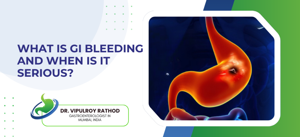
Gastrointestinal bleeding is any bleeding from the digestive tract, anywhere from oesophagus to rectum. It becomes serious when the bleed is fast enough to cause haemodynamic changes, when haemoglobin drops significantly, when symptoms of hypovolaemia develop, or when the suspected source is a high-risk lesion like a peptic ulcer with a visible vessel or oesophageal varices. The location, the rate, and the underlying cause together determine urgency, and any one of those in isolation can underestimate or overestimate the situation.
According to Dr. Vipulroy Rathod, Gastroenterologist in Mumbai, “Patients misjudge GI bleeding in both directions. Some panic over a streak of red blood from a haemorrhoid that’s never dangerous. Others ignore black tarry stools for weeks, which is much more concerning. Melena means significant upper GI bleeding. The colour matters. The volume matters. The symptoms matter. All three together tell the story, and any one in isolation can mislead.”
What GI bleeding looks like clinically?
GI bleeding presents differently depending on where it’s coming from and how fast it’s happening. Recognising the pattern is the first step in working out how urgent things are.
- Upper GI bleeding. Originates above the ligament of Treitz. Oesophagus, stomach, or duodenum. Presented as haematemesis, fresh red or coffee-ground vomitus. Or as melena, black tarry stools from digested blood passing through the small bowel. Occasionally as fresh red blood per rectum if the bleed is massive and rapid.
- Lower GI bleeding originates below the ligament, from the small bowel, colon, or rectum. It usually appears as haematochezia, fresh or maroon blood mixed with stool, sometimes with clots. Common causes include diverticular disease, haemorrhoids, anal fissures, colorectal cancer, and inflammatory bowel disease.
- Occult bleeding. Slow, ongoing blood loss that the patient never sees. Shows up as iron deficiency anemia on routine bloods, sometimes with a positive faecal occult blood test. Often the only sign of a small but persistently bleeding lesion that needs endoscopic investigation.
- Obscure GI bleeding is the term used when upper and lower endoscopy have both come back negative but bleeding continues. The small bowel is usually the source. Investigation needs capsule endoscopy, push enteroscopy, or CT angiography depending on whether the bleed is overt or obscure.
The colour and pattern usually narrow the location down before any test is done. Advanced GI procedures then confirm the source and treat it in the same session in most cases, with endoscopic clips, cautery, band ligation, or injection therapy depending on the lesion.
When GI bleeding becomes serious?
Most GI bleeding episodes are minor and self-limiting. The ones that aren’t tend to declare themselves through specific clinical features.
- Haemodynamic compromise is the clearest marker. Tachycardia. Low blood pressure. Dizziness on standing. Sometimes frank shock. These signs reflect significant volume loss, often before the patient has appreciated how much bleeding has actually occurred. Resuscitation comes first in these cases, with diagnosis following once the patient is stable.
- Melena or haematemesis both indicate upper GI bleeding that has reached a volume significant enough to be obvious to the patient. Coffee-ground vomiting suggests slower or older bleeding, fresh red haematemesis suggests active brisk bleeding, and melena typically means at least 50 to 100 mL of blood has been lost into the upper tract.
- Bleeding in patients with known risk factors changes the threshold completely. Cirrhosis with portal hypertension raises the risk of variceal bleeding, which carries high mortality without urgent endoscopic intervention. Patients on anticoagulants, antiplatelet agents, or NSAIDs have higher bleed rates and worse outcomes when bleeding does occur. A first episode in any of these groups warrants admission regardless of how stable the patient looks initially.
Recurrent bleeding matters even when individual episodes seem minor. Multiple small bleeds point to an underlying lesion that hasn’t been identified, and the cumulative effect on haemoglobin and quality of life justifies investigation rather than continuing to manage each episode separately. Read more on endoscopic treatment for GI bleeding for how modern endoscopic techniques manage most significant bleeds without surgical intervention.
Why choose Dr. Vipulroy Rathod for GI bleeding assessment?
Dr. Vipulroy Rathod has been managing acute and chronic GI bleeding at Fortis Hospital Mulund for over three decades, with the urgent endoscopy capability that serious cases need. Patients arrive at various stages. Some after a single concerning episode. Others after months of intermittent bleeding that’s been managed symptomatically without anyone identifying the source. Both groups need proper assessment, often with endoscopy as the next step rather than another round of empirical iron or PPI treatment.
The judgment that matters most in GI bleeding is timing. Endoscopy too early in a stable patient with self-limiting bleeding adds risk without changing outcomes. Endoscopy too late in a patient with ongoing bleeding allows the haemoglobin to drop further and complications to develop. Getting that balance right is what experience teaches.
Book your consultation today with one of India’s most experienced specialists for GI bleeding assessment and endoscopic management.
Frequently Asked Questions
Not always. A small streak on toilet paper or surface of stool is usually haemorrhoids or an anal fissure. Heavier red bleeding mixed with stool, recurrent episodes, or any bleeding alongside weight loss, change in bowel habit, or anaemia needs investigation regardless of how it looks.
Melena. Which is significant upper GI bleeding. The black colour comes from digested blood that has passed through the small intestine. Usually means at least 50 to 100 mL has been lost into the upper tract, and it shouldn’t be managed at home.
Yes, many episodes do. Self-limiting bleeds from small ulcers, haemorrhoids, or minor inflammation often resolve without intervention. The question isn’t only whether it stops, but whether the source has been identified, because recurrence is common when the underlying lesion remains untreated.
If you’re vomiting blood. If your stools are black and tarry. If there’s heavy red rectal bleeding with clots. If dizziness or fainting accompanies bleeding of any kind. If you have known cirrhosis and any GI bleeding. If you’re on anticoagulants and bleeding has started. Any of these warrant immediate hospital assessment.
Reference links-
- GI Bleeding Management Guidelines, American College of Gastroenterology — https://gi.org/guideline/management-of-patients-with-acute-lower-gastrointestinal-bleeding/
- Variceal and Non-variceal Upper GI Bleeding Standards, World Gastroenterology Organisation — https://www.worldgastroenterology.org/guidelines
