Dr. Vipulroy Rathod

Author name: Dr. Rathod Medical Foundation

What is enteroscopy and how is it different from colonoscopy?

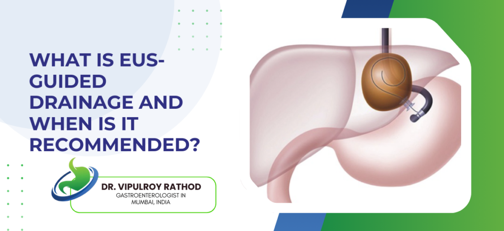
Enteroscopy looks at the small intestine. That’s the part of the gut neither a standard endoscopy nor a colonoscopy can actually reach. Different scope, different purpose, and the patients who need it are usually the ones who’ve already had everything else done and still don’t have answers. According to Dr. VipulRoy, “Double Balloon Endoscopy lets us reach and treat problems in the small bowel that would otherwise go undiagnosed for years. Most patients who need it have already had normal colonoscopy and endoscopy results. That’s exactly when enteroscopy becomes the next step.” What Enteroscopy Actually Is? Most of the small bowel is invisible to standard scopes — Upper endoscopy gets through roughly the first 30 centimetres from the mouth. Colonoscopy comes in from the other end and covers the large intestine. The remaining 6 to 7 metres of small bowel sits between them, untouched by either. Enteroscopy gets in there using a balloon system — A double balloon or single balloon scope uses an overtube and alternating balloon inflation to fold the small intestine back onto itself. The scope edges forward bit by bit. It’s slow, deliberate work — nothing like a routine endoscopy. Treatment happens in the same sitting — Whatever the doctor finds, whether it’s a bleeding vessel, a polyp, or a stricture, it gets dealt with then and there. Biopsy, cauterisation, dilation — all possible without booking a second procedure. It takes time and preparation — Longer than any standard scope. More sedation. More setup. But for patients who’ve been bounced between normal results for months or years, it’s often the only investigation that actually finds something. As a gastroenterology specialist in Mumbai, Dr. Rathod does this at Fortis Hospital Mulund for exactly those cases. How It Differs From Colonoscopy? Colonoscopy is for the large bowel — Colon, rectum, and the very last bit of small bowel called the terminal ileum. That’s its territory. Good for colorectal cancer screening, polyps, IBD. Most people over 50 have had one. It has nothing to do with the small intestine proper. Enteroscopy is for what colonoscopy can’t reach — The jejunum and ileum. The long middle stretch of digestive tract that, before enteroscopy existed, surgeons had to open the abdomen to examine. Nobody did it unless they absolutely had to. They get ordered for completely different reasons — Persistent unexplained GI bleeding after normal upper and lower scopes. Crohn’s suspected in the small bowel. A mass or narrowing picked up on imaging but not reachable by standard endoscopy. Malabsorption that celiac testing didn’t explain. None of these are colonoscopy cases. Ordering the wrong one wastes months — Patients sometimes wait for a colonoscopy when what they actually need is an enteroscopy. The results come back normal, nothing changes, and the actual problem keeps going undiagnosed. You can explore our previous blog What Is EUS-Guided Drainage and When Is It Recommended? to understand how specialised GI procedures fill diagnostic gaps that routine investigations leave behind. Why Choose Dr. Vipulroy Rathod for Enteroscopy in Mumbai? Most GI centres don’t have the equipment for double balloon enteroscopy. The ones that do don’t all have someone who’s been doing it long enough to handle difficult anatomy or unexpected findings mid-procedure. Dr. Vipulroy Rathod has been at Fortis Hospital Mulund for over 30 years doing exactly this kind of advanced diagnostic work. The patients who end up here are often the ones carrying a thick file of inconclusive reports. Sometimes enteroscopy is the first investigation that actually shows something. That happens more often than it should which is why getting the right specialist matters. Still getting normal results but something is clearly wrong? A specialist evaluation of the small bowel may be what’s missing. Book Appointment Call now Frequently Asked Questions Is enteroscopy painful? Done under sedation so there’s no pain during the procedure. Some abdominal discomfort and bloating after is normal and resolves within a day. How long does enteroscopy take?  Typically 60 to 90 minutes depending on how far into the small bowel the doctor needs to go. Recovery adds another couple of hours before discharge. Can enteroscopy treat as well as diagnose? Yes. Bleeding sources can be cauterised, polyps removed, and strictures dilated during the same procedure. It’s not just diagnostic. Who needs enteroscopy instead of colonoscopy? Anyone with unexplained GI bleeding, suspected small bowel Crohn’s, or small bowel tumours where upper and lower endoscopy came back normal. Colonoscopy simply doesn’t reach far enough for these cases. Reference links- Enteroscopy and Small Bowel Endoscopy — American Society for Gastrointestinal Endoscopy Small Intestine Disorders and Diagnosis — American College of Gastroenterology Double Balloon Enteroscopy Clinical Data — National Library of Medicine Small Bowel Endoscopy Standards — World Gastroenterology Organisation

What is enteroscopy and how is it different from colonoscopy? Read More »

What Is EUS-Guided Drainage and When Is It Recommended

EUS-guided drainage is a non-surgical procedure that drains fluid collections pancreatic cysts, abscesses, blocked ducts using a needle guided by real-time ultrasound from inside the gut. No incisions. No surgical entry point. The endoscope does it all. According to Dr. VipulRoy, “Endoscopic Ultrasound guided drainage changed how we handle pancreatic complications. What used to mean surgery or waiting weeks now gets sorted in a single session. Patients recover faster and the risk is far lower.” How EUS-Guided Drainage Works? Endoscope goes in through the mouth — A flexible scope with an ultrasound probe at the tip is guided down into the stomach. It sits right against the gut wall, directly next to the fluid collection on the other side. Doctor sees everything before touching it — The ultrasound shows the cyst or abscess in real time. Size, location, what’s inside. Nothing gets punctured until the image is clear. Needle drains the collection directly — Guided by that live image, a needle passes through the gut wall into the fluid. It drains out in the same session. As a gastroenterology specialist in Mumbai, Dr. Rathod has been performing EUS procedures since 1998, among the first in South Asia to use it clinically. Stent placed if needed — In some cases a small stent goes in to keep the drainage pathway open while the collection fully resolves over the following weeks. No wound, no stitches — Nothing is cut from outside. Most patients leave within 48 hours and recover in days, not weeks. When Is It Actually Recommended? Pancreatic pseudocysts after pancreatitis — Small ones sometimes resolve on their own. Larger ones causing pain, blocking the stomach, or showing infection need drainage. EUS handles it directly without surgery. Walled-off pancreatic necrosis — Dead pancreatic tissue enclosed in a fluid cavity. Used to mean open surgery every time. At specialist centres now, EUS-guided necrosectomy is the standard and outcomes are significantly better. Blocked bile duct when ERCP fails — If conventional ERCP can’t reach the bile duct, EUS creates an alternative route through the stomach wall instead. No surgical incision needed. Deep abscesses difficult to reach from outside — For collections that percutaneous drainage can’t access accurately, EUS sometimes gets there more reliably from inside the gut. You can explore our previous blog Advanced GI Procedures Without Open Surgery in India to understand where EUS-guided drainage fits within the broader picture of minimally invasive GI treatment. Why Choose Dr. Vipulroy Rathod for EUS-Guided Drainage in Mumbai? EUS-guided drainage is not something most gastroenterologists do. It needs both advanced endoscopy skills and real familiarity with pancreatic and biliary complications together. Dr. Vipulroy Rathod has been doing this at Fortis Hospital Mulund since 1998. Patients arrive with complex collections after incomplete management elsewhere. Three decades of this specific work changes what’s possible on difficult cases. Dealing with a pancreatic cyst or abscess? Talk to a specialist before it gets complicated. Book Appointment Call now Frequently Asked Questions Is EUS-guided drainage painful? No. It’s done under sedation or general anesthesia. Most patients feel nothing during the procedure and say recovery was easier than they expected. How long does recovery take?  Most patients go home within 48 hours. Normal activity resumes within a few days. There’s no surgical wound to heal from. Can EUS-guided drainage replace surgery for pancreatic cysts? For most pseudocysts and walled-off necrosis, yes. Surgery is rarely needed now when an experienced endoscopist handles it at the right time. What happens if a stent is placed during the procedure? It keeps the drainage pathway open while the collection resolves. A follow-up endoscopy removes it once imaging confirms healing. Straightforward procedure done under light sedation. Reference links- EUS-Guided Drainage Clinical Overview — American Society for Gastrointestinal Endoscopy Pancreatic Cyst and Pseudocyst Management — American College of Gastroenterology EUS-Guided Biliary and Pancreatic Drainage — National Library of Medicine Interventional EUS Standards — World Gastroenterology Organisation

What Is EUS-Guided Drainage and When Is It Recommended Read More »

Advanced GI Procedures Without Open Surgery in India

A lot of GI conditions that needed open surgery ten or fifteen years ago don’t need it anymore. The endoscope goes in through the mouth or a small access point, reaches the problem, and handles it often in a single sitting. No large wound. Hospital stay measured in hours, not weeks. According to Dr. VipulRoy, “In the last two decades, Advanced Endoscopic Procedures for Complex GI Conditions have replaced open surgery for a large number of patients. The outcomes hold up. But most patients don’t know these options exist until someone tells them.” What Procedures Are Now Done Without Open Surgery? More than most people realise. Here is what’s actually available at a proper therapeutic endoscopy centre. Endoscopic Ultrasound with drainage — Pancreatic cysts, abscesses, fluid collections after pancreatitis. All of these used to sit for weeks before a surgeon would touch them. Endoscopic Ultrasound guided drainage handles it far earlier with a needle placed under real-time imaging. Nothing cut from outside the body. ERCP for bile duct problems — Stones stuck in the bile duct, ducts narrowed by scarring or tumour, bile leaks after gallbladder removal. ERCP reaches all of it from inside the gut. Stones come out. Stents go in. As a gastroenterology specialist in Mumbai, Dr. Rathod has been doing this for over 30 years — including cases referred after failed attempts elsewhere. POEM for achalasia — The overactive muscle blocking the esophagus gets cut through a tunnel built inside the esophageal wall itself. Patients eat normally within a week. No scar. Most don’t miss more than two days of work. Endoscopic Submucosal Dissection — Early tumours and large polyps in the stomach, esophagus, or colon removed by cutting under the mucosal layer. What used to mean bowel resection surgery now means an overnight stay in most cases. Third Space Endoscopy — The broader category that includes POEM and ESD. Working inside the GI wall layers, not just on the surface. Technically demanding. Only a handful of centres in India do it at any real volume. Who Actually Needs These and When? These aren’t for every GI complaint. But for the right conditions, nothing else comes close. Blocked bile ducts — Jaundice with fever and right-sided abdominal pain usually means a stone in the bile duct. That combination moves fast toward sepsis if left alone. ERCP clears it without a surgical incision and most patients feel dramatically better within 24 hours. Pancreatic collections after pancreatitis — Fluid that builds up after a bad episode of pancreatitis used to wait for surgery. EUS-guided drainage gets to it sooner and with far less trauma for someone who is already unwell. Achalasia after dilation stops working — Balloon dilation and Botox injections buy time. When they stop holding, POEM is where most of these patients end up. Surgery is rarely needed anymore for this condition. Early GI cancers found on surveillance — Caught before they’ve spread, tumours in the esophagus and stomach can be removed entirely through the endoscope. You can explore our previous blog What Happens During an Endoscopy Step by Step? to understand how these findings are first picked up during a routine examination. Why Choose Dr. Vipulroy Rathod for Advanced GI Procedures in Mumbai? Most hospitals in India offer basic endoscopy. Third-space procedures, EUS-guided drainage, complex ERCP that’s a much shorter list. Dr. Vipulroy Rathod was among the first in South Asia to use EUS clinically, starting in the late 1990s when most centres here hadn’t heard of it. Three decades later he is still doing it at Fortis Hospital Mulund, including cases that came in after incomplete procedures at other centres. If you have been told surgery is your only option for a GI condition, that assessment is worth a second look. Have a GI condition you were told needs surgery? Get a second opinion from a specialist first. Book Appointment Call now Frequently Asked Questions Are endoscopic GI procedures safe without surgery? Yes. Complication rates are low when performed by an experienced endoscopist with high procedure volume. Most patients are discharged within 24 to 48 hours with minimal post-procedure discomfort. How long is recovery after advanced endoscopic GI procedures? Most patients go home within 24 to 48 hours. A few days of restricted activity at home is typical. Open surgery for the same conditions usually means 5 to 10 days in hospital and several weeks before full recovery. Are these procedures available across India or only in Mumbai? Advanced procedures like POEM, ESD, and EUS-guided drainage are available only at select centres with specialist training. Mumbai has one of the highest concentrations of experienced therapeutic endoscopists in India. Can endoscopic procedures replace surgery for GI cancers? For early-stage GI cancers detected before any spread, endoscopic resection through ESD can be curative. Advanced or metastatic cancers still require surgery or combined treatment depending on the specific case and staging. Reference links- Advanced Endoscopic Procedures Overview — American Society for Gastrointestinal Endoscopy Minimally Invasive GI Treatment Guidelines — American College of Gastroenterology Endoscopic Submucosal Dissection and POEM Data — National Library of Medicine Global Standards in Therapeutic Endoscopy — World Gastroenterology Organisation

Advanced GI Procedures Without Open Surgery in India Read More »

What Happens During an Endoscopy Step by Step?

An endoscopy is a direct visual examination of the digestive tract using a thin, flexible tube with a camera at the tip. No surgical cuts. No general anesthesia in most cases. The procedure gives the doctor a real-time view of what’s happening inside your esophagus, stomach, or intestine that no external scan can match. According to Dr. VipulRoy, “Endoscopy is often the only way to see what’s actually happening inside. Scans tell you something is there. Endoscopy tells you exactly what it is and sometimes lets us treat it in the same sitting.” He explains that Endoscopic Treatment for GERD and Complex GI Conditions allows diagnosis and treatment to happen in a single procedure, which is what makes it so valuable for patients who have been dealing with unresolved GI symptoms for a long time. What Happens Before and During the Procedure? Most patients are more anxious about endoscopy than they need to be. The procedure itself is well-tolerated. Here is exactly what happens from the moment you arrive. Fasting beforehand is mandatory — You will be asked to avoid food and water for at least 6 to 8 hours before the procedure. This clears the stomach so the doctor gets a clean, unobstructed view of the lining. Anything left inside reduces visibility and increases risk. A sedative or throat spray is given — For upper endoscopy, a local anesthetic throat spray numbs the gag reflex. Light sedation is offered in most cases. You remain conscious but relaxed. Most patients remember very little of the procedure itself. The endoscope is passed through the mouth — A thin flexible tube roughly the diameter of a finger is guided down the throat. It passes through the esophagus into the stomach and then the first part of the small intestine if needed. As a gastroenterology specialist in Mumbai, Dr. Rathod performs this with precision to minimize discomfort at every stage. The doctor examines the lining in real time — High-definition images from the camera at the tip appear on a monitor. The doctor looks for inflammation, ulcers, polyps, bleeding sources, or abnormal tissue. Everything is documented as the scope moves through. Biopsies or treatment happen during the same procedure — If something needs to be sampled or removed, it happens right then. Polyps get taken out. Tissue samples get collected. Bleeding gets controlled. No second procedure needed in most cases. What Happens After the Procedure? Recovery from endoscopy is quick. But knowing what to expect helps. You rest for 20 to 30 minutes after the procedure — The sedation wears off during this time. Nursing staff monitor your vitals. You will feel drowsy but the discomfort is minimal for most patients. Mild bloating or throat soreness is normal — Air is used during the procedure to open up the GI tract for better visibility. Some of that passes out afterward causing temporary bloating. Throat soreness from the scope usually resolves within 24 hours. Results are discussed before you leave — In most cases the doctor reviews findings with you the same day. If biopsies were taken, those results typically come back within a few days. You can explore our previous blog What Is POEM Procedure and Who Performs It in Mumbai? to understand how endoscopic procedures extend beyond diagnosis into advanced therapeutic interventions. You cannot drive yourself home — Sedation affects coordination and judgment for several hours after. Arrange for someone to take you home. Most patients return to normal activity the following day without any restrictions. Why Choose Dr. Vipulroy Rathod for Endoscopy in Mumbai? Endoscopy is only as good as the person performing it. Lesions get missed. Biopsies get taken from the wrong site. Therapeutic interventions get skipped when they shouldn’t be. Dr. Vipulroy Rathod has performed over 30 years of diagnostic and therapeutic endoscopy at Fortis Hospital Mulund, including complex cases referred after incomplete procedures elsewhere. If you need an endoscopy done properly the first time, that experience is what makes the difference. Start Your Treatment Journey Today Book Appointment Call now Frequently Asked Questions Is endoscopy painful? Most patients feel no pain during the procedure. Throat discomfort and mild bloating afterward are common but resolve within 24 hours. How long does an endoscopy take? The procedure itself takes 15 to 30 minutes. With preparation and recovery time, plan for around 2 hours at the facility. Can endoscopy detect cancer? Yes. Endoscopy can identify suspicious tissue and collect biopsies for lab analysis. Early detection through endoscopy significantly improves treatment outcomes. Do I need someone to accompany me for an endoscopy? Yes. Sedation affects your ability to drive and make decisions for several hours after the procedure. Arrange for an adult to take you home. Reference links- Endoscopy Overview and Indications — American Society for Gastrointestinal Endoscopy Upper GI Endoscopy Guidelines — American College of Gastroenterology Endoscopy Procedure and Safety — National Library of Medicine Digestive Endoscopy Standards — World Gastroenterology Organisation

What Happens During an Endoscopy Step by Step? Read More »

What Is POEM Procedure and Who Performs It in Mumbai?

POEM Peroral Endoscopic Myotomy is used to treat achalasia and certain spastic esophageal conditions. No incisions on the body. No surgical entry points. The entire procedure goes through the mouth with a flexible endoscope. According to Dr. VipulRoy, “POEM works by cutting the muscle that’s preventing food from reaching the stomach. There’s no wound to recover from. Patients who are good candidates see real, lasting improvement.” For patients with achalasia or swallowing problems that haven’t responded to other treatments, Advanced Endoscopic Procedures for Complex GI Conditions explains how these cases are handled without surgery. What Actually Happens During a POEM Procedure? People hear “through the mouth” and think it’s straightforward. It’s not. POEM is a third-space endoscopic procedure. The work happens inside the wall of the esophagus, not just on its surface. A small cut is made in the inner esophageal lining — The endoscope goes in through the mouth. That cut opens access to a space between the layers of the esophageal wall. Nothing on the outside of the body is involved at any point. A tunnel is created down toward the stomach — Through that internal space, the doctor works toward the lower esophageal sphincter. That’s the muscle that refuses to relax in achalasia. It’s what’s been stopping food from getting through. The muscle gets cut under direct view — The myotomy happens here. Muscle fibers are divided carefully, with the endoscope showing exactly what’s being cut. For a deeper understanding of this level of endoscopic work, visit Third Space Endoscopy in Mumbai to see how these procedures are approached as a gastroenterology specialist in Mumbai. The entry point is clipped shut — After the myotomy, the tunnel opening is closed with endoscopic clips. No stitches. No external wound. Most patients leave the hospital within 48 hours. When Is POEM the Right Call? Not every swallowing issue ends up here. But for some conditions, there’s genuinely nothing that works as well. Achalasia cardia — This is what POEM was built for. The sphincter at the bottom of the esophagus stops relaxing. Food piles up. Patients often say it feels like something is stuck in the chest after every meal. POEM fixes the actual problem, not just the symptoms. Type II achalasia — Responds particularly well. Studies consistently put success above 90% at two years. Surgery was the standard before POEM existed. For most patients with this subtype, it’s no longer necessary. Spastic esophageal disorders — Diffuse esophageal spasm and similar conditions cause chest pain that gets mistaken for cardiac problems. POEM goes after the muscle dysfunction directly rather than working around it. When earlier treatments have failed — Balloon dilation or Botox that worked for a while and then didn’t. POEM is where a lot of these cases end up. You can explore our previous blog What Is ERCP and When Is It Recommended? to understand how similar biliary and GI procedures are evaluated and recommended. Why Choose Dr. Vipulroy Rathod for POEM in Mumbai? POEM sits at the more demanding end of therapeutic endoscopy. Not many gastroenterology doctors in Mumbai do it regularly, and fewer still have three decades of third-space endoscopic work behind them. Dr. Vipulroy Rathod has managed cases that came in after failed procedures elsewhere or after patients were told open surgery was their only remaining option. That isn’t always true. But figuring out whether it applies to your specific case requires an honest evaluation from someone who actually knows the procedure from the inside. Start Your Treatment Journey Today Book Appointment Call now Frequently Asked Questions Is POEM better than surgery for achalasia? For most patients, yes. No external cuts, faster recovery, and outcomes that hold up well compared to Heller myotomy over time. How long does the POEM procedure take? Around 60 to 90 minutes under general anesthesia. Most patients are home within 48 hours and eating normally within a week. Can POEM still be done after balloon dilation? Yes. A previous dilation doesn’t rule it out. POEM is still a viable option even when earlier treatments have stopped working. What risks come with POEM? Mucosal tears, gas-related discomfort, and acid reflux afterward are the ones that come up most. An endoscopist in Mumbai who performs this regularly keeps those risks low. Reference links- POEM Procedure and Achalasia Management — American Society for Gastrointestinal Endoscopy Achalasia Diagnosis and Treatment Guidelines — American College of Gastroenterology Peroral Endoscopic Myotomy Clinical Data — National Library of Medicine Esophageal Motility Disorders — World Gastroenterology Organisation

What Is POEM Procedure and Who Performs It in Mumbai? Read More »

Stent After Gallbladder Surgery

Gallbladder surgery, commonly known as cholecystectomy, is performed to remove the gallbladder due to conditions like gallstones, inflammation, or infection. This procedure is generally safe and effective, helping relieve symptoms caused by gallbladder dysfunction. However, in some cases, complications can arise following the surgery, particularly involving the bile ducts. To address these issues, a stent after gallbladder surgery may be necessary. Dr. Vipulroy Rathod, a globally recognized gastroenterologist in Mumbai, India, explains: “A stent helps ensure proper bile flow from the liver to the small intestine, preventing obstructions or leaks that can occur post-surgery. A well-placed stent can significantly improve recovery, reduce the risk of infection, and help prevent bile duct complications after gallbladder removal.” Dr. Rathod specializes in advanced gastrointestinal procedures, including gallbladder stent surgery. His expertise ensures that the procedure is performed with precision and minimal discomfort, promoting optimal recovery. Dr. Vipulroy Rathod’s approach to post-surgery care focuses on reducing risks and providing tailored solutions to help patients recover quickly and without complications. First, let’s explore how a stent works and its role following gallbladder surgery. What Is a Stent and Why Is It Used After Gallbladder Surgery? A stent is a small, flexible tube placed into a duct or vessel to help keep it open. After gallbladder removal, a cholecystectomy stent may be used if there’s a risk of bile duct obstruction or narrowing. The bile duct, which carries bile from the liver to the small intestine, may become blocked or constricted due to surgery or other complications. A stent helps maintain an open passage, ensuring bile continues to flow properly and preventing further issues such as jaundice or infections. Now, let’s discuss the situations in which a stent becomes essential after gallbladder surgery. When Is a Stent Needed After Gallbladder Surgery? A stent is typically needed if there are complications such as: Bile duct obstruction Caused by scar tissue or injury during the surgery. Bile leakage If bile leaks from the duct into surrounding tissues, a stent can help redirect bile flow. Strictures Narrowing of the bile duct that may occur after surgery. Infection Bile duct infections can lead to blockages that require stent placement for proper drainage. Dr. Vipulroy Rathod emphasizes that a stent is an effective solution for managing these complications and improving healing. If you’re experiencing any of these issues after gallbladder surgery, a stent may be necessary. Speak to a specialist for expert guidance. Book Appointment Call now How do you know when a stent might be required after gallbladder surgery? Let’s discuss the signs to look out for. Symptoms Indicating the Need for a Stent Post-Surgery Symptoms that may indicate the need for a stent after gallbladder removal include: Yellowing of the skin and eyes (jaundice): A sign of bile flow disruption. Severe abdominal pain: Often in the upper right quadrant, which can indicate bile duct issues. Nausea and vomiting: Persistent nausea after surgery may signal a bile duct blockage. Fever: Associated with infection or bile duct obstruction. If these symptoms occur, Dr. Rathod recommends early intervention to prevent complications and ensure a smooth recovery. What is the process for placing a stent after gallbladder removal? Let’s break down the procedure. How Is a Stent Placed After Gallbladder Removal? 1. Preparation The patient is usually under anesthesia or sedation so as to comfort him or her throughout the process. 2. Endoscopic Procedure The stent can be installed endoscopically, with the help of ERCP (Endoscopic Retrograde Cholangiopancreatography), which is a procedure that implies inserting an endoscope through the mouth. 3. Accessing the Bile Duct The endoscope will be inserted into the digestive tract to the bile duct where the stent will be placed. 4. Stent Insertion A thin, flexible tube (the stent) is then carefully placed in the bile duct in order to keep the passage open so that bile can flow through. 5. Placement Verification The position of the stent is checked using imaging techniques like X-ray or fluoroscopy to ensure it’s properly placed. 6. Post-Procedure Monitoring After placement, the patient is monitored for any signs of discomfort, infection, or complications. This minimally invasive approach helps prevent further complications and ensures bile continues to flow properly after gallbladder removal. What should you do after the stent is placed to ensure proper care? Let’s explore the essential steps for maintaining the stent after surgery. Post-Surgery Care and Maintenance of a Stent Following the placement of a stent, there are certain aftercare guidelines that one has to adhere to: Periodic examinations To check the position of a stent and there are no blockages. Watch for infection Watch for any signs of infection, e.g. fever or abnormal pain. Light lifting Do not engage in hard work until your doctor clears you. Hydration and diet Drink as much as possible of fluids and eat a healthy diet to aid in the healing process. Dr. Rathod stresses that post-care must be consistent to avoid complications such as displacement of stents or infection. Can you avoid the need for a stent after gallbladder surgery? Here’s how preventive measures can help. How to Prevent the Need for a Stent After Gallbladder Surgery Although certain factors such as scarring are unavoidable, the risk of the necessity of stenting could be minimized by: Early removal of gallstones Before surgery, it will avoid complications such as inflammation or infection. Choosing an experienced surgeon Lowers the risks of bile duct injury during surgery. Post-surgery follow-up The early identification of issues with the bile duct can avoid the necessity of a stent. Maintaining a healthy lifestyle Reducing factors that contribute to bile duct issues, such as obesity or excessive alcohol consumption. Dr. Rathod emphasizes the importance of proactive healthcare and regular follow-ups for better long-term outcomes. Conclusion The placement of a stent after gallbladder surgery can be a crucial step in ensuring proper bile flow and preventing complications such as infection or obstruction. While it is often necessary to address post-surgery issues, proper care and early intervention can help manage the risks

Stent After Gallbladder Surgery Read More »

Thinning of the Stomach Lining: Causes, Symptoms, and Treatment

The stomach lining, also known as the mucosal lining, acts as a protective barrier that shields the stomach walls from strong digestive acids and enzymes. This lining produces mucus and other protective substances that prevent irritation while supporting proper digestion. However, as this protective layer thins, the stomach becomes more vulnerable to inflammation, ulcers, and digestive discomfort. This condition, commonly referred to as thinning of the stomach lining, can develop due to infections, long-term medication use, autoimmune conditions, or chronic inflammation. According to Dr. Vipulroy Rathod, an internationally acclaimed gastroenterologist in Mumbai, India, “The stomach’s mucosal lining plays a critical role in digestive health. When it weakens or thins, early diagnosis and proper care become essential to prevent complications.” He further adds that maintaining stomach health through timely treatment can significantly improve long-term digestive wellbeing. With over three decades of experience in diagnosing and managing complex gastrointestinal conditions, Dr. Vipulroy Rathod is an expert in advanced endoscopic techniques and the management of digestive diseases. He has helped thousands of patients regain digestive health through precise diagnosis and personalized treatment plans. His experience in evaluating and managing thinning of the stomach lining allows patients to receive expert care, accurate assessments, and effective treatment strategies tailored to their condition. What factors can weaken the stomach’s protective barrier? Let’s explore the common causes behind thinning of the stomach lining. What Causes Thinning of the Stomach Lining? Several medical and lifestyle factors can contribute to damage or thinning of the stomach lining. Understanding these causes helps identify the condition early and prevent further damage. Common causes include: Helicobacter pylori infection A bacterial infection that causes chronic inflammation and gradually damages the stomach’s mucosal lining. Autoimmune gastritis A condition where the immune system mistakenly attacks cells of the stomach lining. Long-term use of medications Frequent use of painkiller medication including NSAIDs can irritate and weaken the stomach lining over time. Chronic inflammation Persistent irritation in stomach can slowly reduce the protective thickness of the mucosal lining. Alcohol abuse Alcohol can damage the stomach lining and increase inflammation. Aging Natural aging can sometimes contribute to the gradual thinning of stomach tissues. Nutritional deficiencies Deficiency of vitamin B12 or other nutrients may have an impact on stomach health. Concerned about your digestive health? Connect with a specialist to better understand your stomach condition and explore the right treatment approach. Book Appointment Call now Could your digestive symptoms be linked to stomach lining damage? Let’s discuss the signs that may indicate thinning of the stomach lining. Symptoms of Thinning Stomach Lining Symptoms can develop over time and vary from mild pain to more apparent digestive issues. Common symptoms include: Persistent stomach pain or discomfort Burning sensation in the upper abdomen Bloating or frequent indigestion Loss of appetite Nausea or vomiting Fatigue or weakness due to nutrient deficiencies. Unexplained weight loss in some cases. Symptoms of vitamin B12 deficiency such as fatigue or nerve problems. These symptoms can be similar to other gastrointestinal conditions and hence proper assessment should be carried out for correct diagnosis. Now, let’s discover the diagnostic methods used to evaluate this condition. How is Thinning of the Stomach Lining Diagnosed? Upper gastrointestinal endoscopy The stomach is visually analyzed with the help of a flexible camera and abnormalities are identified. Read More Biopsy during endoscopy To evaluate inflammation or thinning of mucosal lining, a small tissue sample can be taken. Read More Blood tests A blood test is done to test vitamin deficiencies, infections or autoimmune markers. Read More H. pylori testing Breath, stool or biopsy tests are used to identify bacterial infections. Read More Imaging tests In certain instances, imaging can be suggested to assess digestive health. Read More Early diagnosis plays a vital role in preventing further damage to the stomach lining and improving treatment outcomes. How can the thinning of the stomach lining be treated effectively? Let’s explore the treatment approaches that help restore stomach health. Treatment Options for Thinning of the Stomach Lining Treatment aims at addressing the underlying cause, protecting stomach lining and improving digestion. The most common treatment methods are: Acid-Reducing Medications A physician can prescribe proton pump inhibitors (PPIs) or H2 blockers to reduce stomach acid and protect weakened stomach wall from further harm. H. pylori Infection Treatment In case of bacterial infection like the Helicobacter pylori, the combination of antibiotics and acid-reducing drugs is used to eliminate the infection. Nutritional Supplements and Vitamin Deficiencies may occur in patients whose stomach lining is too thin especially of vitamin B12, iron or folate. Supplements aid in the restoration of nutrient levels and general digestive wellbeing. Anti-Inflammatory and Protective Medications Certain medications help coat and protect the stomach lining, reducing inflammation and allowing the mucosal lining to heal. Dietary Modifications Irritation can be reduced by a stomach-friendly diet that avoids spicy foods, alcohol, caffeine, and processed foods. Lifestyle Changes Reducing alcohol intake, quitting smoking, stress management and proper weight can contribute a great deal to stomach health. Regular Monitoring and Follow-Up In some instances, endoscopic or medical assessment after every few months can be recommended to check the status of the stomach lining and to ascertain that it is healing well. Early treatment and proper management play an important role in protecting the stomach lining and preventing complications.Looking for effective treatment options? Speaking with a specialist can help identify the best approach for your condition. Book Appointment Call now Can the thinning of the stomach lining be prevented from worsening? Let’s explore simple yet effective preventive steps. How to Prevent Further Damage to the Stomach Lining Preventive measures focus on protecting the stomach lining and maintaining a healthy digestive system. Preventive measures can be helpful and these are: Eating a healthy and balanced diet rich in vitamins and nutrients Limiting alcohol intake to reduce stomach irritation Avoiding unnecessary use of painkillers that harm the mucosal lining Managing stress, which can worsen digestive issues Regular medical check-ups for individuals with persistent digestive symptoms Treating infections early, especially H. pylori Protecting the stomach’s mucosal lining helps reduce the risk of complications

Thinning of the Stomach Lining: Causes, Symptoms, and Treatment Read More »

What Is ERCP and When Is It Recommended?

ERCP  Endoscopic Retrograde Cholangiopancreatography is a procedure that accesses the bile duct and pancreatic duct from inside the digestive tract. No surgical incision. No separate imaging appointment. Diagnosis and treatment happen in the same sitting. According to Dr. VipulRoy, “Pancreatic and Biliary Stone Extraction through ERCP lets us remove stones, place stents, and take tissue samples all in one procedure. Patients who would have needed surgery ten years ago walk out the next morning.” What ERCP Actually Does Inside Your Body? Here’s what most people picture. Something complicated and scary. But ERCP is minimally invasive. Done under sedation.  It diagnoses blockages in real time Bile duct stones, strictures, and tumour-related blockages get visualised directly during ERCP. No guessing from external scans. The doctor sees exactly what’s causing your problem and where. Stones get removed in the same session Biliary stones blocking your bile duct get extracted right there during the procedure. No surgery needed. Most patients feel dramatically better within 24 hours of stone removal. Stents get placed for strictures and blockages Narrowed bile ducts from cancer, inflammation, or surgical scarring get opened with stent placement during ERCP. That restores bile flow and relieves jaundice faster than any alternative approach. Tissue sampling happens during the procedure Suspicious areas inside bile ducts get biopsied through ERCP. Confirmed tissue diagnosis without surgery means faster treatment decisions and significantly less risk for you overall. Sphincterotomy treats sphincter dysfunction When the sphincter controlling bile duct drainage is too tight ERCP cuts it precisely. That simple intervention resolves recurrent biliary pain that medication alone never adequately fixes.Know more about how EUS works for your specific situation at Gastroenterology specialist in mumbai. When Does Your Doctor Actually Recommend ERCP? Not every bile duct problem needs ERCP. But certain situations make it the most effective and least invasive option available. Honestly, knowing when it’s the right call takes genuine clinical experience. Bile duct stones causing jaundice or infection Jaundice, fever, and right upper abdominal pain together often signal a bile duct stone. That combination needs ERCP urgently. Waiting makes things significantly worse very quickly. Blocked bile ducts from pancreatic cancer Pancreatic cancer frequently blocks the bile duct as it grows. ERCP places a stent to restore drainage and relieve jaundice while the oncology treatment plan gets finalised for you. Chronic pancreatitis with ductal complications Pancreatic duct strictures and stones causing recurrent pain get treated through ERCP. Surgery used to be the only option for these patients. Now it often isn’t. Post-surgical bile duct complications Bile leaks or strictures after gallbladder or liver surgery get managed effectively through ERCP without reoperation. That’s a genuinely significant advantage for patients already recovering from surgery. For more on advanced biliary endoscopy expertise, read our previous blog on What Conditions Can EUS Diagnose. Why Choose Dr. VipulRoy Rathod for ERCP? Dr. Vipulroy Rathod has spent three decades performing ERCP on some of the most complex biliary cases in India. Patients arrive after failed procedures elsewhere or after being told surgery is their only remaining option. And honestly? That’s exactly where his 30 years of focused biliary endoscopy experience makes the biggest difference. Don’t let a blocked bile duct sit untreated because you haven’t found the right specialist yet. Think you could benefit from specialized care? Reach out today to explore your treatment options. Book Appointment Call now Frequently Asked Questions Is ERCP painful and how long does recovery take? It’s done under sedation so there’s no pain during the procedure. Most patients go home the next day feeling significantly better already. Can ERCP fail and what happens then?  In difficult anatomy cases ERCP occasionally can’t access the duct. EUS-guided drainage then offers an alternative route without surgery. How is ERCP different from a regular endoscopy? Regular endoscopy examines the gut surface. ERCP goes inside your bile and pancreatic ducts to diagnose and treat duct-specific problems directly. Is ERCP safe for elderly or high-risk patients? Yes. Its minimally invasive nature actually makes it safer than surgery for elderly and medically complex patients who can’t tolerate general anaesthesia well. References links ERCP Overview and Indications — American Society for Gastrointestinal Endoscopy Biliary Stone Management — American College of Gastroenterology ERCP Clinical Guidelines — National Library of Medicine Pancreatic and Biliary Disease — World Gastroenterology Organisation

What Is ERCP and When Is It Recommended? Read More »

What Conditions Can EUS Diagnose?

EUS (Endoscopic Ultrasound) puts an ultrasound probe inside the digestive tract, right next to the organ being examined. That proximity changes what’s visible. Structures that look unclear on a CT scan or MRI show up in far more detail from 2 to 3 centimetres away. According to Dr. VipulRoy, “Endoscopic Ultrasound gives us images that external scans simply can’t match for certain organs. The pancreas, bile duct, and surrounding lymph nodes — EUS sees all of it from the inside. For patients who’ve had inconclusive CT results, this is often where the answer finally comes.” Pancreatic and Biliary Conditions EUS Diagnoses The pancreas and bile duct are where EUS genuinely shines brightest. These structures sit deep inside your body where external imaging struggles badly. EUS reaches them from inside your stomach wall and delivers clarity nothing else consistently matches. Pancreatic cancer and early tumours Small pancreatic masses invisible on CT get picked up through EUS. Pancreatic cancer treatment outcomes depend heavily on how early the tumour gets found. EUS finds it early when it still matters most. Pancreatic cysts and precancerous changes Not all cysts are benign. EUS classifies exactly what type of cyst you have and whether it carries malignant potential. That classification changes your entire management plan completely. Bile duct stones hiding from ultrasound Common bile duct stones that regular ultrasound misses regularly show up clearly on EUS. Patients with repeated normal scans but ongoing symptoms finally get their answer here. Biliary strictures and cholangiocarcinoma Narrowing of the bile duct and suspected bile duct cancer get evaluated with a precision that no external scan delivers. Tissue sampling happens in the same session through EUS-guided biopsy. Chronic pancreatitis and ductal changes Pancreatitis treatment planning depends on understanding ductal anatomy accurately. EUS maps pancreatic duct changes with a detail level that MRI and CT simply can’t match for your specific situation. Know more about how EUS works for your specific situation at Gastroenterology specialist in mumbai. GI Tract and Cancer Staging Conditions EUS Diagnoses Beyond the pancreas EUS evaluates your entire digestive tract wall layer by layer. That capability makes it the gold standard for GI cancer staging and submucosal lesion assessment. And honestly, that staging accuracy changes surgical decisions more than most patients realise. Oesophageal and gastric cancer staging Tumour depth and lymph node involvement get assessed precisely through EUS before surgery. Your surgeon needs that information to make the right call about what operation you actually need. Rectal cancer staging GI cancer treatment for rectal tumours depends on accurate local staging. EUS shows exactly how deep the tumour goes and whether nearby nodes are involved. That shapes chemotherapy and surgical planning entirely. Submucosal tumours beneath the gut lining GISTs, lipomas, and carcinoid tumours hiding beneath the surface get identified and characterised through EUS. Standard endoscopy sees a bulge. EUS tells you exactly what that bulge actually is. Mediastinal lymph node assessment Enlarged lymph nodes in your chest get sampled through EUS-guided biopsy from inside the oesophagus. No chest surgery. No CT-guided biopsy. Just precise minimally invasive tissue diagnosis. For more on how EUS differs from standard endoscopy, read our previous blog on What Is EUS and How Does It Differ from Endoscopy. Why Choose Dr. Vipul Roy Rathod for EUS Diagnosis? Dr. Vipulroy Rathod has spent over 25 years building a diagnostic accuracy in EUS that directly changes patient outcomes. India’s first FASGE fellow. Over 30 years of advanced endoscopy practice. Patients arrive after months of inconclusive scans and leave with real answers. Because EUS in the right hands doesn’t just find conditions. It finds them early enough to actually do something about them. Frequently Asked Questions Can EUS diagnose conditions that CT and MRI already missed? Yes. EUS regularly finds pancreatic lesions, bile duct stones, and submucosal tumours that CT and MRI couldn’t identify clearly. Does EUS only diagnose or can it treat conditions too?  EUS does both. Diagnosis and interventional treatment like drainage or biopsy often happen during the same single procedure. How many conditions can be evaluated in one EUS session? Multiple structures get assessed in one session. Pancreas, bile duct, lymph nodes, and gut wall layers all get evaluated without repeat procedures. Is EUS recommended before cancer surgery for staging? Absolutely. EUS staging before GI cancer surgery gives your surgical team precise information that directly determines which operation is the right one for you. References links EUS Diagnostic Applications — American Society for Gastrointestinal Endoscopy EUS in GI Cancer Staging — National Library of Medicine Pancreatic and Biliary EUS Guidelines — American College of Gastroenterology Endoscopic Ultrasound Overview — World Gastroenterology Organisation

What Conditions Can EUS Diagnose? Read More »

What Is EUS and How Does It Differ from Endoscopy?

EUS Endoscopic Ultrasound combines a standard endoscope with an ultrasound probe at the tip. It goes into the digestive tract the same way a regular endoscopy does. But instead of just looking at the surface, it images the layers beneath and the organs sitting right outside the gut wall. According to Dr. VipulRoy, “Endoscopic Ultrasound is not an upgraded endoscopy. It’s a different investigation entirely. Endoscopy tells you what the lining looks like. EUS tells you what’s happening underneath it and in the organs immediately behind it.” What Standard Endoscopy Actually Does? Regular endoscopy is incredibly useful. Don’t get that wrong. But it has a clear limit. It shows your doctor the inner lining of your digestive tract. That’s it. What’s beyond that lining stays invisible. It examines the surface lining only Standard endoscopy spots ulcers, inflammation, bleeding, and surface polyps well. But anything sitting beneath the gut wall or in nearby organs simply doesn’t show up at all. It’s the right tool for many common conditions Colonoscopy and upper GI endoscopy diagnose GERD, gastritis, IBD, and colorectal polyps effectively. For surface-level problems endoscopy is exactly what you need. Biopsies from the surface are possible Tissue samples from the gut lining get taken during standard endoscopy. But deeper lesions sitting beneath the surface or in the pancreas? That requires something more powerful entirely. It can’t evaluate your pancreas or bile duct This is the biggest limitation. The pancreas and bile duct sit behind and beside the gut wall. Standard endoscopy can’t reach or image them with any real diagnostic clarity for you. Know more about how EUS works for your specific situation at Gastroenterology specialist in mumbai. What EUS Does That Endoscopy Simply Can’t? EUS combines a flexible endoscope with a high-frequency ultrasound probe at its tip. It goes inside your digestive tract. Then it uses ultrasound to see through the wall and into surrounding structures. That combination is genuinely powerful. It images the pancreas from inside your stomach wall The pancreas is notoriously difficult to see from outside the body. EUS reaches it directly from inside and gives image clarity that no CT scan or MRI consistently matches for early lesions. It evaluates all layers of your gut wall Endoscopic Ultrasound shows every individual layer of your digestive tract wall. Submucosal tumours hiding beneath the surface get identified and assessed precisely here. EUS-guided biopsy reaches deep tissue Tissue samples from the pancreas, lymph nodes, and bile duct get collected through EUS-guided fine needle aspiration. No surgery. No incisions. Just accurate targeted sampling exactly where it’s needed. Cancer staging happens in real time Tumour depth, vascular involvement, and lymph node status get assessed during the same EUS session. Your oncologist gets the precise information they need immediately from one focused procedure. For more on why EUS expertise changes diagnostic outcomes, read our previous blog on Best Gastroenterologist in Mumbai for Pancreatic Cancer Diagnosis. Why Choose Dr. VipulRoy Rathod for EUS in Mumbai? Dr. Vipulroy Rathod has spent over 25 years building a level of EUS expertise that most gastroenterologists in India simply haven’t developed. He’s India’s first FASGE fellow. He’s trained doctors from 35 countries. And honestly? Patients who arrive after months of inconclusive standard endoscopy results leave with real answers after one properly performed EUS session. That’s what the right specialist actually does for you. Think you could benefit from specialized care? Reach out today to explore your treatment options. Book Appointment Call now Frequently Asked Questions Can EUS completely replace standard endoscopy for diagnosis? No. Both serve different purposes. Standard endoscopy handles surface conditions well. EUS goes deeper for complex cases that endoscopy simply can’t evaluate properly. Is EUS more uncomfortable than regular endoscopy? Both are done under sedation. Most patients feel no difference in comfort level between the two procedures at all. When does a doctor recommend EUS over standard endoscopy?  When symptoms suggest pancreatic, biliary, or submucosal problems that regular endoscopy can’t evaluate clearly enough for a confident diagnosis. How long does an EUS procedure take compared to endoscopy?  Standard endoscopy takes around 15 to 20 minutes. EUS takes 30 to 45 minutes depending on complexity of your specific case. References links Endoscopic Ultrasound Overview — American Society for Gastrointestinal Endoscopy EUS vs Standard Endoscopy — National Library of Medicine Diagnostic Endoscopy Guidelines — American College of Gastroenterology Advanced Endoscopy Clinical Overview — World Gastroenterology Organisation

What Is EUS and How Does It Differ from Endoscopy? Read More »

Scroll to Top
Call Now Button