

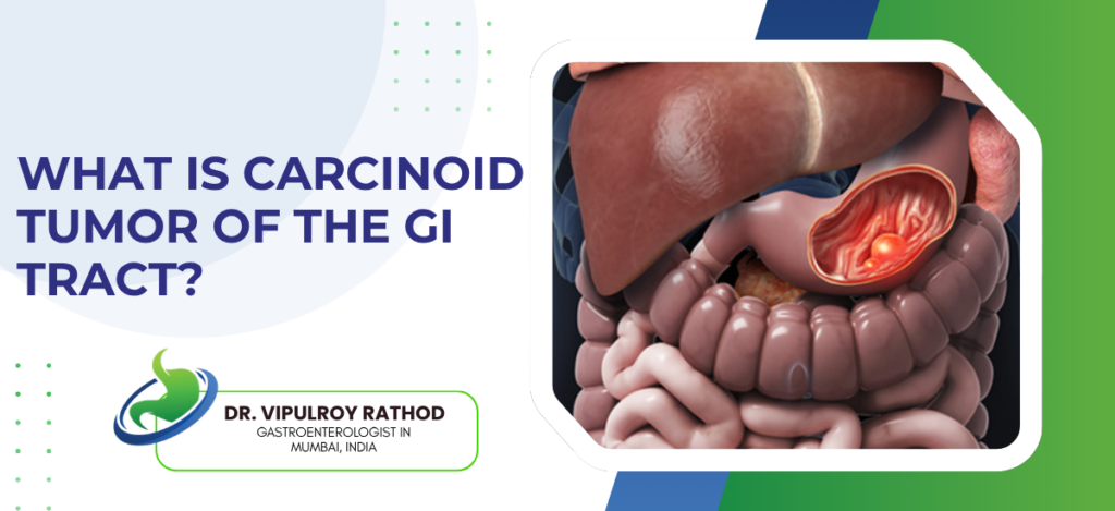
A carcinoid tumor is a slow-growing neuroendocrine tumor that arises from the hormone-producing cells lining the digestive tract, most commonly in the small intestine, rectum, stomach, or appendix. Many are found by chance during an endoscopy done for something else, since they often cause no symptoms for years. When they’re small and caught early, they can frequently be removed endoscopically without open surgery.
According to Dr. Vipulroy Rathod, Gastroenterologist in Mumbai, “Carcinoid tumors are among the more reassuring things we find, provided they’re caught early. Many are picked up incidentally as a small nodule during a routine scope, and when they’re limited to the surface layers we can often remove them endoscopically. The picture changes if they’ve grown or spread, which is exactly why finding them early matters so much.”
What Are the Symptoms and How Are They Found?
The tricky part is that carcinoid tumors are often silent, which shapes how they get diagnosed.
Most cause no symptoms at all. A large proportion are discovered by accident as a small nodule during an endoscopy or surgery performed for an unrelated reason, long before they would ever have announced themselves.
Larger or spread tumors can produce carcinoid syndrome. Flushing, diarrhoea, wheezing, and eventually heart effects appear when the tumor releases enough hormones into the bloodstream, though this happens mostly once the disease has reached the liver.
Endoscopy with biopsy is the starting point. Direct visualisation finds the nodule and a biopsy confirms its neuroendocrine nature, which is often the moment an incidental lesion is first identified for what it is.
EUS then assesses the depth. Endoscopic ultrasound shows how far into the bowel wall the tumor extends and whether nearby lymph nodes are involved, which is the information that decides whether endoscopic removal is safe or surgery is needed.
Getting that depth assessment right is exactly what a specialist in endoscopic submucosal dissection uses to plan whether a carcinoid can be removed through the scope.
How Are GI Carcinoid Tumors Treated?
Treatment turns almost entirely on the tumor’s size, depth, and whether it has spread.
Small, superficial carcinoids often come out endoscopically. For tumors confined to the mucosa or submucosa and under a certain size, EMR or ESD removes them completely through the scope with no abdominal incision.
Larger or deeper tumors need surgery. Once a carcinoid has grown beyond what endoscopic resection can safely clear, or reached the muscle layer, surgical removal of the affected segment becomes the appropriate route.
Spread disease brings in additional therapies. When the tumor has reached lymph nodes or the liver, hormone-blocking drugs like somatostatin analogues and targeted treatments help control both symptoms and progression.
Follow-up matters because these are slow but persistent. Carcinoids can recur or progress over years, so surveillance with endoscopy, imaging, and hormone markers continues well after the initial treatment.
Because some carcinoids first announce themselves through bleeding rather than hormones, knowing how such lesions are managed helps. Read more on GI bleeding treatment to understand how bleeding GI lesions are controlled endoscopically without surgery.
Why Choose Dr. Vipulroy Rathod for GI Tumors?
Dr. Vipulroy Rathod has been diagnosing and treating GI neuroendocrine and other tumors at Fortis Hospital Mulund for over 30 years, with more than 20,000 EUS procedures giving him the depth assessment that decides whether a carcinoid can be removed endoscopically. Small tumors that other centres send to surgery are frequently removed here through the scope. Physicians from 35 countries have trained under his approach at Fortis.
Book your consultation today with one of India’s most experienced specialists for GI carcinoid tumor diagnosis and endoscopic treatment.
Frequently Asked Questions
It is a slow-growing neuroendocrine tumor arising from hormone-producing cells in the digestive tract, most often in the small intestine, rectum, stomach, or appendix.
Many cause no symptoms and are found incidentally. Larger or spread tumors can cause flushing, diarrhoea, abdominal pain, and, in carcinoid syndrome, wheezing and heart effects.
Diagnosis uses endoscopy with biopsy, endoscopic ultrasound to assess depth, imaging, and blood or urine tests for hormones like chromogranin A and 5-HIAA.
Small localised carcinoids are often removed endoscopically by EMR or ESD, while larger or spread tumors need surgery and sometimes hormone-blocking or targeted therapy.
Reference
- Gastroenteropancreatic Neuroendocrine Tumours: Diagnosis and Management — NCBI/PMC, https://www.ncbi.nlm.nih.gov/pmc/articles/PMC5836830/
- Endoscopic Diagnosis and Treatment of Gastrointestinal Neuroendocrine Tumours — NCBI/PMC, https://www.ncbi.nlm.nih.gov/pmc/articles/PMC6182296/
