 Skip to content
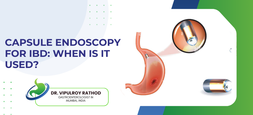
Colonoscopy reaches the colon and terminal ileum well, but a large stretch of small bowel between those two points stays largely invisible to it. The gap capsule endoscopy fills a camera the size of a pill that the patient swallows, which transmits images of the entire small bowel to a recorder worn around the waist. In IBD, particularly Crohn’s disease, it’s this segment that gets missed most often and investigated last, which is exactly where the capsule changes the clinical picture.
According to Dr. Vipulroy Rathod, Gastroenterologist in Mumbai, “Capsule endoscopy picks up small bowel Crohn’s disease that colonoscopy and CT scans have both missed. Patients who’ve had unexplained anemia, weight loss, or abdominal pain for months with normal previous investigations often get a clear answer from a capsule study that nobody thought to do earlier.”
When Is Capsule Endoscopy Used in IBD?
Four situations in IBD practice consistently push the clinical decision toward a capsule study.
Suspected small bowel Crohn’s with negative colonoscopy is the clearest indication. Up to 30% of Crohn’s patients have disease limited to the small bowel, and the capsule reaches those segments with a diagnostic yield that beats both CT and barium studies.
Monitoring mucosal healing after treatment matters more now that deep remission is the target in Crohn’s management, and the capsule gives a direct mucosal view of segments that no external imaging assesses with enough resolution to count.
Obscure GI bleeding in a patient with known IBD often points toward small bowel involvement, and when upper and lower endoscopy come back clean, the capsule is the next logical step rather than another round of cross-sectional imaging.
Assessing post-operative recurrence after bowel resection in Crohn’s disease, particularly at the neo-terminal ileum, is a setting where the capsule adds detail that colonoscopy alone doesn’t provide for segments proximal to the anastomosis.
These decisions sit within the broader framework of endoscopic IBD management, and a specialist in IBD management weighs which investigation fits the patient’s specific presentation.
What Are the Limitations and Risks?
Capsule endoscopy is powerful but not without conditions that limit its use.
Capsule retention is the main risk. If the bowel is narrowed by a stricture, the capsule can get stuck, and a patency capsule or cross-sectional imaging beforehand checks whether the passage is clear enough to proceed safely.
The capsule cannot take biopsies or treat what it finds, so any suspicious lesion it identifies still needs a follow-up procedure to sample tissue or deliver therapy.
Bowel preparation affects image quality, and an inadequately prepared small bowel produces images that are harder to read and easier to misinterpret, making patient adherence to the prep protocol essential.
Not every patient swallows it easily. Those with known oesophageal strictures or severe dysphagia sometimes need the capsule placed endoscopically rather than swallowed, which adds a step but doesn’t change the imaging itself.
Understanding how colonoscopy fits alongside the capsule for IBD diagnosis helps clarify which tool does what. Read more on Crohn’s detection to understand what colonoscopy finds in Crohn’s disease and where it hands over to the capsule.
Why Choose Dr. Vipulroy Rathod for Capsule Endoscopy?
Dr. Vipulroy Rathod has been using capsule endoscopy as part of his IBD and small bowel practice at Fortis Hospital Mulund for over two decades, and the patients who reach him have often already had colonoscopy and CT without a clear answer. That experience means the clinical picture gets read correctly, the right preparatory steps are taken, and the capsule findings are interpreted in context rather than in isolation. Physicians from 35 countries have trained under his approach at Fortis.
Book your consultation today with one of India’s most experienced specialists for capsule endoscopy and IBD assessment.
Frequently Asked Questions
A pill-sized camera swallowed by the patient that travels through the digestive tract and transmits small bowel images to a recorder worn around the waist.
It is used when colonoscopy cannot reach suspected small bowel Crohn’s, when monitoring mucosal healing, or when obscure bleeding points to small bowel involvement.
Yes. It detects ulcers, erosions, and inflammatory changes in the small bowel that colonoscopy cannot reach, making it valuable for diagnosing and monitoring small bowel Crohn’s.
The main risk is capsule retention in a narrowed bowel segment, so patients with suspected strictures are assessed with a patency capsule or imaging first.
Reference
- Capsule Endoscopy for Crohn’s Disease: Current Status of Diagnosis and Management — NCBI/PMC, https://www.ncbi.nlm.nih.gov/pmc/articles/PMC4706954/
- Capsule Endoscopy Findings for the Diagnosis of Crohn’s Disease — NCBI/PMC, https://www.ncbi.nlm.nih.gov/pmc/articles/PMC6394710/
