Dr. Vipulroy Rathod

Author name: Dr. Rathod Medical Foundation

Survival Rate of Digestive Cancers in India

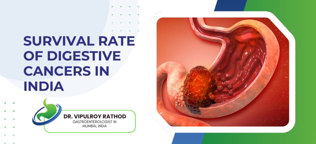
Survival rates for digestive cancers in India vary significantly by cancer type and stage at diagnosis. Colorectal cancer caught at Stage 1 has a 5-year survival rate above 90%, dropping to under 15% at Stage 4. Pancreatic cancer overall 5-year survival sits around 8 to 10% because most cases are found late. Stomach and oesophageal cancers follow a similar pattern. Stage at detection is the single biggest factor. According to Dr. Vipulroy Rathod, an experienced Gastroenterologist in Mumbai, “Survival statistics for digestive cancers in India look poor largely because most patients arrive at advanced stage, and that is not a reflection of how treatable these cancers are when found early, it is a reflection of how late investigation happens.” What Are the Survival Rates for Different Digestive Cancers? Each cancer type has its own survival profile. Some are very treatable early. Some are difficult even at early stage. Worth knowing the difference. Colorectal Cancer, Best Survival Profile: Stage 1 colorectal cancer has 5-year survival above 90% with surgery, Stage 2 around 70 to 80%, Stage 3 drops to 40 to 60% depending on nodal involvement, Stage 4 under 15%  the gap between early and late detection here is bigger than almost any other GI cancer. Stomach Cancer, Dramatically Stage Dependent: Early gastric cancer caught at mucosal level has 5-year survival above 95% with endoscopic resection, but most Indian patients present at Stage 3 or 4 where survival drops to 20 to 30%, and that gap exists because early stomach cancer produces no symptoms that feel alarming. Pancreatic Cancer, Hardest Numbers: Overall 5-year survival around 8 to 10% in India, surgical resection at Stage 1 pushes that to 20 to 30%, but less than 20% of pancreatic cancer cases in India are caught at resectable stage because the investigation that finds it early simply isn’t being done at the right time. Oesophageal Cancer, Tobacco and Late Presentation: 5-year survival for localised oesophageal cancer is around 40 to 50%, for regional spread drops to 20 to 25%, for distant metastasis under 5% and most Indian patients present with dysphagia that’s already been progressing for months before anyone scopes them. Stage at diagnosis changes survival more than any treatment advance in the last decade. Specialist in GI cancer treatment catches cases early enough for those better survival numbers to actually apply. What Actually Determines Survival in Digestive Cancers? Not just stage. Several factors compound each other and most patients aren’t told about all of them. Stage at Diagnosis, Dominates Everything: Already said it but it needs repeating because patients focus on treatment options when the more important variable is already fixed at the point of diagnosis, finding it early is worth more than any specific treatment protocol. Investigation Accuracy Matters: Wrong staging means wrong treatment and wrong treatment wastes time the patient doesn’t have, EUS-based staging for pancreatic, oesophageal, and gastric cancers consistently outperforms CT-only staging and that accuracy difference has direct survival implications. Time Between Suspicion and Diagnosis: Indian data consistently shows months of delay between first symptom and confirmed diagnosis, every month of delay in GI cancers with fast doubling times like pancreatic cancer meaningfully changes what stage the patient arrives at for treatment. Access to the Right Specialist: General physician to gastroenterologist to oncologist referral chain takes time in India and patients with vague symptoms often cycle through multiple consultations before anyone orders the investigation that actually finds something. Survival statistics look discouraging until you look at what they’re measuring. Read more on what EUS can diagnose to understand how the right investigation changes the starting point. Why Choose Dr. Vipulroy Rathod Dr. Vipulroy Rathod 30 years gastroenterology, EUS since 1998, trained physicians from 35 countries. Sees GI cancer cases at every stage at Fortis Hospital Mulund and has been doing this long enough to know that the patients who do well are almost always the ones who got properly investigated before the disease declared itself loudly. Months of normal reports. Vague symptoms nobody pinned down. Most patients with that history leave here with an actual finding. Not a referral. A diagnosis. Start Your Treatment Journey Today Book Appointment Call now Frequently Asked Questions What is the survival rate for pancreatic cancer in India? Overall 5-year survival is around 8 to 10% but rises to 20 to 30% when caught at a surgically resectable early stage. Which digestive cancer has the best survival rate in India? Colorectal cancer caught at Stage 1 has a 5-year survival above 90% making it one of the most survivable GI cancers when detected early. Does early detection really improve digestive cancer survival? Yes, significantly. Stage 1 and Stage 4 survival rates for most digestive cancers differ by 60 to 80 percentage points. Why are digestive cancer survival rates lower in India than in Western countries? Later stage at diagnosis due to delayed investigation and limited routine screening programmes accounts for most of the survival gap. Reference links- GI Cancer Survival Data India — Indian Council of Medical Research Digestive Cancer Outcomes and Staging — World Gastroenterology Organisation

Survival Rate of Digestive Cancers in India Read More »

What Tests Detect Pancreatic Disease Early

Tests used to detect pancreatic disease early include endoscopic ultrasound (EUS), CT scan, MRI with MRCP, blood tests including CA 19-9 and amylase, and endoscopic retrograde cholangiopancreatography (ERCP). EUS is the most sensitive tool for early pancreatic lesions, finding tumours under 2cm that CT and MRI regularly miss. Blood tests alone are not reliable for early detection. According to Dr. Vipulroy Rathod, Gastroenterologist in Mumbai, “Most pancreatic disease gets found late because patients and doctors rely on CT scans that look normal while something small but significant is already present, EUS exists specifically to close that gap and it does it consistently.” Which Tests Are Used to Detect Pancreatic Disease Early? Not all tests detect pancreatic disease equally. The pancreas lies deep, and standard investigations often reach their limits quickly. EUS, the Most Accurate Tool Available: Probe sits millimetres from pancreatic surface inside the stomach wall, images from that proximity find sub-2cm lesions, ductal changes, cysts, and early tumours that external scans miss routinely and that’s not an occasional occurrence, it’s the norm. CT Scan, Good for Obvious Disease: CT is fast, widely available, and picks up larger masses and distant metastasis well, but misses early pancreatic cancer consistently because the organ’s retroperitoneal location means too much tissue between the scanner and the target. MRI with MRCP: Better than CT for ductal anatomy and cystic lesions, MRCP maps the pancreatic duct without contrast injection and is particularly useful for patients with suspected chronic pancreatitis or intraductal papillary mucinous neoplasms where duct changes matter. Blood Tests, Limited but Useful: CA 19-9 elevated in pancreatic cancer but also in benign conditions like pancreatitis and bile duct obstruction, amylase and lipase spike during acute pancreatitis episodes, none of these replace imaging but they help build the clinical picture when used alongside it. Right investigation from the start changes what gets found. Specialists in endoscopic ultrasound don’t just order tests, they know exactly which one applies to the specific clinical picture in front of them. When Should You Get Tested for Pancreatic Disease? Most people wait for a major symptom, but by then the window for early detection has often already passed Family History, Start Now: One first-degree relative with pancreatic cancer means active EUS surveillance should already be happening, not being considered for the future, because the precancerous changes EUS finds are exactly the ones that matter before they become cancer. Chronic Pancreatitis Patients: Repeated pancreatic inflammation carries real malignant transformation risk over time and patients with established chronic pancreatitis need periodic EUS monitoring not just symptom management between flares. New Onset Diabetes After 50: Already covered under risk factors but worth repeating here because it’s the most commonly missed clinical trigger for pancreatic investigation, gets filed as endocrine disease, managed with medication, pancreas never checked. Vague Symptoms, Normal CT: Upper abdominal discomfort, unexplained weight loss, back pain, nausea that doesn’t explain itself and a CT that shows nothing — that combination is exactly the clinical picture where EUS finds things and CT didn’t, not occasionally but regularly. Don’t wait for symptoms to get obvious before investigating properly. Read more on POEM procedure to understand what advanced endoscopic intervention looks like when early detection leads to action. Why Choose Dr. Vipulroy Rathod Dr. Vipulroy Rathod has been doing EUS since 1998. Over 30 years in gastroenterology. Trained physicians from 35 countries. At Fortis Hospital Mulund he handles the full pancreatic disease spectrum from initial investigation through complex intervention and has seen enough normal CT reports with abnormal EUS findings to know exactly why the right test matters. Patients come in after months of reassurance based on one scan. Most leave with a finding nobody else looked for. That’s the difference. Start Your Treatment Journey Today Book Appointment Call now Frequently Asked Questions Can EUS detect pancreatic cancer before symptoms appear? Yes, EUS regularly finds early pancreatic lesions and ductal changes in high-risk patients before any symptoms develop. Is CA 19-9 reliable for early pancreatic cancer detection? No, CA 19-9 is elevated in benign conditions too and is not reliable enough for standalone early detection without imaging. How often should high-risk patients get EUS for pancreatic surveillance? Most guidelines recommend annual EUS surveillance for high-risk patients including those with BRCA2 mutations or strong family history. Does MRCP replace EUS for pancreatic diagnosis? No, MRCP maps ductal anatomy well but EUS provides superior sensitivity for small lesions and allows biopsy in the same session.   Reference links- Pancreatic Disease Diagnosis and Surveillance — American College of Gastroenterology Early Pancreatic Cancer Detection Guidelines — World Gastroenterology Organisation

What Tests Detect Pancreatic Disease Early Read More »

Cancer Staging in Digestive Cancers

Cancer staging in digestive cancers uses the TNM system to classify how far cancer has spread: T for tumour depth into the organ wall, N for lymph node involvement, M for distant metastasis. Stage 1 is localised, Stage 4 means spread to distant organs like liver or lungs. Staging directly decides whether surgery is possible, what treatment sequence applies, and what realistic outcomes look like for each patient. According to Dr. Vipulroy Rathod, an experienced Gastroenterologist in Mumbai, “Most patients arrive with a stage assigned from a CT report done in a hurry and that stage is frequently wrong EUS changes T and N staging in a significant proportion of GI cancer cases and the treatment changes with it.” Why Does Accurate Staging Change Everything? Wrong stage means wrong treatment. Simple as that. Here’s where staging errors actually happen. CT Misses Small Nodal Deposits: Standard CT regularly misses lymph node involvement in early GI cancers because nodes need to be visibly enlarged to show up, and small deposits in normal-sized nodes are exactly what EUS finds and CT doesn’t. T Stage Gets Underestimated on CT: Tumour depth into the organ wall is consistently harder to assess from outside the body, and understaging the T component means patients get offered endoscopic resection for a tumour that has already gone deeper than the scan suggested. Restaging After Treatment Gets Skipped: After chemotherapy or radiation the tumour needs restaging before surgery is reconsidered, this step gets skipped more often than it should and patients go into surgery without anyone confirming what the treatment actually did to the tumour. Stage 4 Gets Missed Early: Small liver metastases and peritoneal deposits are regularly absent on initial staging scans and show up later, which is why high-risk cases need more thorough staging workup not just a single CT before treatment decisions get made. Staging isn’t a one-time checkbox. Read more on POEM procedure to understand how advanced endoscopic procedures work alongside cancer staging in GI management. How Does Diabetes Increase Pancreatic Disease Risk? Alcohol is a Group 1 carcinogen. No safe level for cancer risk has been established and the GI tract takes the most direct hit of any organ system. Liver Cancer Through Cirrhosis: Chronic alcohol use causes cirrhosis and cirrhosis is the strongest single risk factor for hepatocellular carcinoma, cirrhotic patients carry a 1 to 5% annual liver cancer risk regardless of whether they’ve stopped drinking by that point. Colorectal Cancer, Even Moderate Drinking: Risk rises linearly with consumption and even 1 to 2 drinks per day is associated with measurably increased colorectal cancer risk in large population studies, something most patients are genuinely surprised to hear when told directly. Oesophageal Cancer with Smoking Combined: Alcohol and tobacco act synergistically on oesophageal tissue and the combined risk is multiplicative not additive, heavy drinkers who smoke sit in a risk category that justifies regular upper endoscopy surveillance rather than waiting for symptoms to show up. Stomach Cancer Through Mucosal Damage: Alcohol directly damages gastric mucosal lining and chronic exposure creates persistent inflammation that increases H. pylori susceptibility and accelerates the gastritis to cancer progression sequence faster than either factor alone. Both together are worse than either alone and risk doesn’t reset quickly after stopping. Read more on AI in GI endoscopy to understand how modern detection tools are changing early cancer surveillance. Why Choose Dr. Vipulroy Rathod Dr. Vipulroy Rathod has 30 years in gastroenterology, EUS since 1998, trained physicians from 35 countries. Stages GI cancers at Fortis Hospital Mulund with EUS accuracy that CT-dependent workups consistently miss and has seen enough staging errors from outside referrals to know exactly where the gaps are. Patients arrive with a stage. Gets verified here before anyone commits to a treatment plan. That’s the difference between right treatment and expensive wrong treatment. Start Your Treatment Journey Today Book Appointment Call now Frequently Asked Questions What is TNM staging in digestive cancers? TNM describes tumour depth, lymph node involvement, and distant metastasis to classify how far digestive cancer has spread. Is Stage 3 digestive cancer curable? Yes in some cases, Stage 3 cancers with lymph node involvement can still be treated with combined chemotherapy, radiation, and surgery. Why is EUS better than CT for staging GI cancers? EUS images from inside the GI tract giving millimetre-level accuracy for tumour depth and nearby lymph nodes that CT misses regularly. Does staging change during treatment? Yes, restaging after chemotherapy or radiation is standard to assess tumour response before surgery is reconsidered.   Reference links- GI Cancer Staging Guidelines — American College of Gastroenterology Digestive Cancer TNM Staging — World Gastroenterology Organisation

Cancer Staging in Digestive Cancers Read More »

Scroll to Top
Call Now Button