Patient Profile
Patient Name: Savitaben (name changed for confidentiality)
Age: 61 | Gender: Female | Location: Pune, Maharashtra
Referred By: Oncologist — unexplained weight loss workup
Tests: CT abdomen: normal | MRI pancreas: normal | CA 19-9: mildly elevated
Complaint: 7 kg unexplained weight loss over 4 months, back pain, mild jaundice
Patient Background:
Savitaben, a 61-year-old from Pune, Maharashtra, visited Dr. Vipulroy Rathod at Fortis Hospital Mulund after four months of unexplained weight loss that had left her physician and oncologist without answers. Like many patients with early pancreatic pathology, Savitaben’s symptoms of weight loss, back pain, and newly developing jaundice were deeply concerning, yet both a CT abdomen and MRI pancreas had returned completely normal results.
With her CA 19-9 mildly elevated and her condition visibly worsening, her oncologist recognised that standard imaging had reached its limits. Knowing that EUS can detect lesions that CT and MRI miss entirely, she referred Savitaben urgently to the best Gastroenterologist Mumbai Dr. Vipulroy Rathod, pioneer of interventional EUS in South Asia with over 20,000 procedures to his name.
Symptoms
- Unexplained weight loss — 7 kg over four months with no dietary cause
- Back pain — persistent, dull mid-back discomfort
- Mild jaundice — new onset yellowing of skin and eyes
- Fatigue — progressive generalised weakness and low energy
Diagnostic Method
- CT abdomen — performed at referring hospital; reported normal, no mass identified
- MRI pancreas — detailed pancreatic imaging; also reported normal
- CA 19-9 blood marker — mildly elevated, initially attributed to age
- Endoscopic Ultrasound (EUS) + EUS-FNA — high-resolution imaging from within the GI tract, with same-session fine needle aspiration for tissue diagnosis
Disease Diagnosed
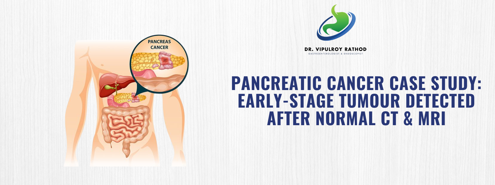
As a leading gastroenterology doctor in Mumbai, Dr. Vipulroy Rathod identified a 1.4 cm hypoechoic mass in the pancreatic head — precisely the area both prior scans had cleared. EUS-guided fine needle aspiration (EUS-FNA) confirmed the diagnosis: pancreatic ductal adenocarcinoma, detected at a surgically resectable stage.
Why Standard Imaging Missed It:
- CT and MRI regularly miss lesions under 2 cm, particularly in the pancreatic head near the bile duct
- EUS operates from within the GI tract, offering proximity and resolution no external scan can replicate
Lesions of 5–7 mm are detectable with EUS far beyond the capability of CT or MRI
Treatment Plan
As the most experienced GI specialist in Mumbai for advanced pancreatic diagnosis, Dr. Vipulroy Rathod performed a linear EUS with EUS-guided fine needle aspiration (EUS-FNA) under conscious sedation locating, imaging, and biopsying the mass in a single 40-minute session.
Why EUS-FNA Was the Right Investigation
1. Unmatched Resolution EUS places the ultrasound probe millimetres from the pancreas, detecting lesions CT and MRI cannot see.
2. Same-Session Tissue Diagnosis A 22G needle was passed through the scope wall under real-time guidance with no separate biopsy procedure required.
3. Caught at a Resectable Stage At 1.4 cm, the tumour was still surgically operable. A delay of even 3 months would likely have closed that window.
4. Zero Additional Hospital Stay Performed under conscious sedation, Savitaben was discharged the same day.
How the Procedure Was Performed
- Savitaben was placed under conscious sedation with no general anaesthesia required.
- A linear array echoendoscope was advanced to the duodenum, adjacent to the pancreatic head.
- A 1.4 cm hypoechoic mass was identified as invisible on both prior CT and MRI scans.
- A 22G FNA needle was passed through the scope wall into the lesion under real-time ultrasound guidance.
- Three passes were made, withdrawing cytological samples sufficient for a definitive tissue diagnosis.
Procedure Summary
- Procedure: Linear EUS + EUS-Guided Fine Needle Aspiration (EUS-FNA)
- Finding:4 cm pancreatic head mass — missed by CT and MRI
- Duration: 40 minutes | Anaesthesia: Conscious sedation
- Hospital Stay: Same-day discharge
“This case is one that stays with me. Two normal scans, and the cancer was already there. Any patient with unexplained weight loss, back pain, or rising CA 19-9 even with a normal CT deserves an EUS evaluation by a specialist. We do this every single day.” Dr. Vipulroy Rathod, FASGE | Gastroenterology Specialist in Mumbai | Visiting Professor, Yale University
Post-Surgery Guidelines
- Soft diet for 24 hours following the procedure
- Monitor for any post-FNA fever, pain, or bleeding and report immediately
- Oncology and surgical referral within 48 hours of cytology confirmation
- Pancreatic cancer treatment planning to begin immediately upon confirmed diagnosis
- All follow-up imaging and surgical coordination managed through Fortis Hospital Mulund
Outcome
| Timepoint | Result |
|---|---|
| During EUS | 1.4 cm mass identified in pancreatic head — missed by CT and MRI |
| Same Session | EUS-FNA performed; cytology samples obtained in 3 passes |
| 48 Hours | Cytology confirmed pancreatic ductal adenocarcinoma |
| 2 Weeks | Savitaben underwent Whipple procedure at Fortis Hospital Mulund |
| Post-Surgery | Tumour successfully resected at early, operable stage |
| Follow-up | Curative treatment pathway opened — surgery would not have been possible 3 months later |
Long-Term Expectations With dietary modifications and alcohol avoidance, Rajan’s prognosis is excellent. Annual follow-up with Dr. Rathod will monitor pancreatic function and digestive health long-term.
Patient Feedback
“Every scan said I was normal. I knew something was wrong, but nobody could find it. Dr. Rathod found it in 40 minutes. He didn’t just make a diagnosis — he gave me a chance at treatment. — Savitaben
Experiencing unexplained symptoms with normal scan results? Book a consultation with Dr. Vipulroy Rathod today.